Structure-function insights into the yeast Dam1 kinetochore complex
- PMID: 19889968
- PMCID: PMC2773187
- DOI: 10.1242/jcs.004689
Structure-function insights into the yeast Dam1 kinetochore complex
Abstract
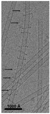
Faithful segregation of genetic material during cell division requires the dynamic but robust attachment of chromosomes to spindle microtubules during all stages of mitosis. This regulated attachment occurs at kinetochores, which are complex protein organelles that are essential for cell survival and genome integrity. In budding yeast, in which a single microtubule attaches per kinetochore, a heterodecamer known as the Dam1 complex (or DASH complex) is required for proper chromosome segregation. Recent years have seen a burst of structural and biophysical data concerning this interesting complex, which has caught the attention of the mitosis research field. In vitro, the Dam1 complex interacts directly with tubulin and self-assembles into ring structures around the microtubule surface. The ring is capable of tracking with depolymerizing ends, and a model has been proposed whereby the circular geometry of the oligomeric Dam1 complex allows it to couple the depolymerization of microtubules to processive chromosome movement in the absence of any additional energy source. Although it is attractive and simple, several important aspects of this model remain controversial. Additionally, the generality of the Dam1 mechanism has been questioned owing to the fact that there are no obvious Dam1 homologs beyond fungi. In this Commentary, we discuss recent structure-function studies of this intriguing complex.
Figures


Similar articles
-
Molecular architecture of the Dam1 complex-microtubule interaction.Open Biol. 2016 Mar;6(3):150237. doi: 10.1098/rsob.150237. Open Biol. 2016. PMID: 26962051 Free PMC article.
-
The Dam1 kinetochore complex harnesses microtubule dynamics to produce force and movement.Proc Natl Acad Sci U S A. 2006 Jun 27;103(26):9873-8. doi: 10.1073/pnas.0602249103. Epub 2006 Jun 15. Proc Natl Acad Sci U S A. 2006. PMID: 16777964 Free PMC article.
-
Structural view of the yeast Dam1 complex, a ring-shaped molecular coupler for the dynamic microtubule end.Essays Biochem. 2020 Sep 4;64(2):359-370. doi: 10.1042/EBC20190079. Essays Biochem. 2020. PMID: 32579171 Free PMC article. Review.
-
Subunit organization in the Dam1 kinetochore complex and its ring around microtubules.Mol Biol Cell. 2011 Nov;22(22):4335-42. doi: 10.1091/mbc.E11-07-0659. Epub 2011 Sep 30. Mol Biol Cell. 2011. PMID: 21965284 Free PMC article.
-
Kinetochore-microtubule interactions in chromosome segregation: lessons from yeast and mammalian cells.Biochem J. 2017 Oct 18;474(21):3559-3577. doi: 10.1042/BCJ20170518. Biochem J. 2017. PMID: 29046344 Review.
Cited by
-
Centromeric heterochromatin: the primordial segregation machine.Annu Rev Genet. 2014;48:457-84. doi: 10.1146/annurev-genet-120213-092033. Epub 2014 Sep 18. Annu Rev Genet. 2014. PMID: 25251850 Free PMC article. Review.
-
The essentiality of the fungus-specific Dam1 complex is correlated with a one-kinetochore-one-microtubule interaction present throughout the cell cycle, independent of the nature of a centromere.Eukaryot Cell. 2011 Oct;10(10):1295-305. doi: 10.1128/EC.05093-11. Epub 2011 May 13. Eukaryot Cell. 2011. PMID: 21571923 Free PMC article.
-
A TOG Protein Confers Tension Sensitivity to Kinetochore-Microtubule Attachments.Cell. 2016 Jun 2;165(6):1428-1439. doi: 10.1016/j.cell.2016.04.030. Epub 2016 May 5. Cell. 2016. PMID: 27156448 Free PMC article.
-
The Opposing Functions of Protein Kinases and Phosphatases in Chromosome Bipolar Attachment.Int J Mol Sci. 2019 Dec 7;20(24):6182. doi: 10.3390/ijms20246182. Int J Mol Sci. 2019. PMID: 31817904 Free PMC article. Review.
-
Amphitelic orientation of centromeres at metaphase I is an important feature for univalent-dependent meiotic nonreduction.J Genet. 2014 Aug;93(2):531-4. doi: 10.1007/s12041-014-0393-9. J Genet. 2014. PMID: 25189254 No abstract available.
References
-
- Cheeseman, I. M. and Desai, A. (2008). Molecular architecture of the kinetochore-microtubule interface. Nat. Rev Mol. Cell. Biol. 9, 33-46. - PubMed
-
- Cheeseman, I. M., Anderson, S., Jwa, M., Green, E. M., Kang, J., Yates, J. R., 3rd, Chan, C. S., Drubin, D. G. and Barnes, G. (2002). Phospho-regulation of kinetochore-microtubule attachments by the Aurora kinase Ipl1p. Cell 111, 163-172. - PubMed
Publication types
MeSH terms
Substances
Grants and funding
LinkOut - more resources
Full Text Sources
Molecular Biology Databases

