. 2009 Nov 17;106(46):19575-80.
doi: 10.1073/pnas.0905884106. Epub 2009 Nov 4.

Rapid modulation of spine morphology by the 5-HT2A serotonin receptor through kalirin-7 signaling

Affiliations

Rapid modulation of spine morphology by the 5-HT2A serotonin receptor through kalirin-7 signaling

Kelly A Jones et al. Proc Natl Acad Sci U S A. .

Abstract

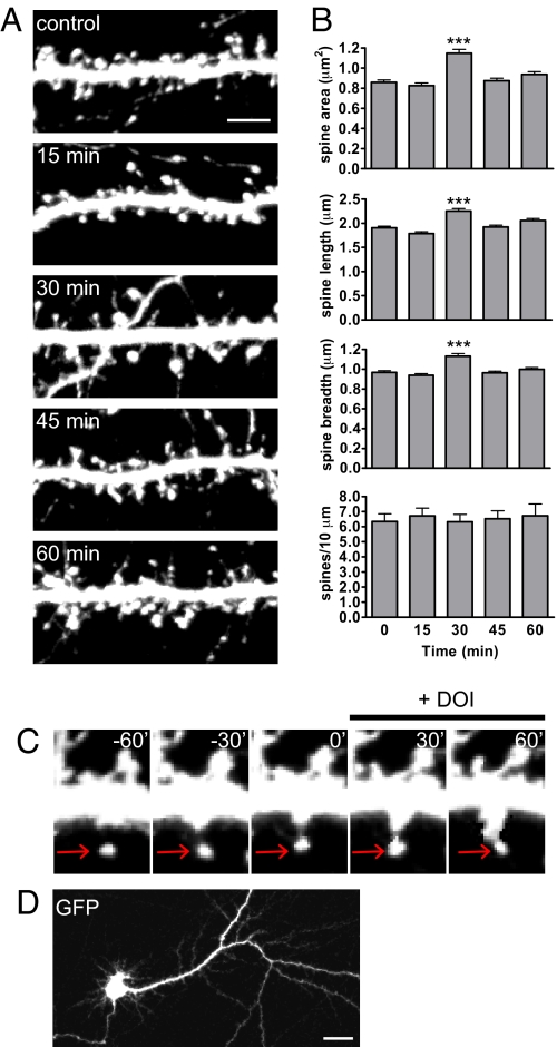
The 5-HT(2A) serotonin receptor is the most abundant serotonin receptor subtype in the cortex and is predominantly expressed in pyramidal neurons. The 5-HT(2A) receptor is a target of several hallucinogens, antipsychotics, anxiolytics, and antidepressants, and it has been associated with several psychiatric disorders, conditions that are also associated with aberrations in dendritic spine morphogenesis. However, the role of 5-HT(2A) receptors in regulating dendritic spine morphogenesis in cortical neurons is unknown. Here we show that the 5-HT(2A) receptor is present in a subset of spines, in addition to dendritic shafts. It colocalizes with PSD-95 and with multiple PDZ protein-1 (MUPP1) in a subset of dendritic spines of rat cortical pyramidal neurons. MUPP1 is enriched in postsynaptic density (PSD) fractions, is targeted to spines in pyramidal neurons, and enhances the localization of 5-HT(2A) receptors to the cell periphery. 5-HT(2A) receptor activation by the 5-HT(2) receptor agonist DOI induced a transient increase in dendritic spine size, as well as phosphorylation of p21-activated kinase (PAK) in cultured cortical neurons. PAK is a downstream target of the neuronal Rac guanine nucleotide exchange factor (RacGEF) kalirin-7 that is important for spine remodeling. Kalirin-7 regulates dendritic spine morphogenesis in neurons but its role in neuromodulator signaling has not been investigated. We show that peptide interference that prevents the localization of kalirin-7 to the postsynaptic density disrupts DOI-induced PAK phosphorylation and spine morphogenesis. These results suggest a potential role for serotonin signaling in modulating spine morphology and kalirin-7's function at cortical synapses.


Conflict of interest statement

The authors declare no conflict of interest.

Figures

Fig. 1.
Fig. 1.
5-HT2A receptors are present in spines and colocalize with synaptic proteins in cultured cortical neurons. (A) Endogenous 5-HT2A receptors localize to the soma and dendrites of mature cultured cortical neurons (DIV 24). (B) Flag-tagged 5-HT2A receptors localize to intracellular endomembranes (“ie”), the dendritic shaft (“ds”), and spine-like structures (“sp”) in the dendrites of mature cultured cortical neurons. (C) Higher magnification of spine-like structures in B. A line scan through the spine and shaft shows increased fluorescence intensity in the head compared to the shaft. (D) Co-expression of flag-5-HT2A and GFP reveal localization of 5-HT2A receptors to dendritic spines (“sp”) and the dendritic shaft (“ds”). (E) Endogenous 5-HT2A receptors colocalize with the presynaptic marker bassoon in the dendrites of cultured cortical neurons (arrowheads). (F) Endogenous 5-HT2A receptors colocalize with PSD-95 in the dendrites of cultured cortical neurons (arrowheads). (G) Histogram of PSD-95 puncta that colocalize with 5-HT2A receptors. (H) 5-HT2A receptors localize to smaller spines. GFP-transfected cells were co-stained for endogenous 5-HT2A receptors. A larger proportion of spines that contained 5-HT2A had small-to-medium area (< 1 μm2) (arrowheads), while fewer large spines (> 1 μm2) contained 5-HT2A signal (open arrowhead). (I) Histogram of the areas of spines that contain 5-HT2A receptors. Insets show typical dendritic spine shapes for each range of spine area: small (area <1 μm2), medium (area = 1 μm2), and large (area >1 μm2). [Scale bar, 5 μm (E, F, and H); 20 μm (A).]
Fig. 2.
Fig. 2.
MUPP1 promotes 5-HT2A receptor membrane trafficking in COS-7 cells. (A) Endogenous 5-HT2A receptors and MUPP1 partially colocalize in mature cultured cortical neurons (DIV 24). (B) MUPP1 overexpressed in COS-7 cells localizes to the cell periphery (“p”), intracellular endomembranes (“ie”), and vesicular-like structures (“v”). (C) 5-HT2A receptors overexpressed in COS-7 cells localize to intracellular endomembranes (“ie”) and vesicular-like structures (“v”). (D) Co-expression of the 5-HT2A receptor and MUPP1 results in increased localization of 5-HT2A receptors to the cell periphery compared to 5-HT2A expression alone. (E) Line scans show increased fluorescence intensity of 5-HT2A at the cell periphery when co-expressed with MUPP1. [Scale bar, 5 μm (A).]
Fig. 3.
Fig. 3.
Activation of 5-HT2A receptors with DOI results in dendritic spine remodeling. (A) Time course of dendritic spine morphogenesis in response to 1 μM DOI. Mature cultured cortical neurons (DIV 21) expressing GFP were treated with 1 μM DOI in ACSF + APV for 0–60 min, and fixed immediately. (B) Quantification of dendritic spine morphogenesis in A. Spine area, length, and breadth are increased after 30 min DOI, while spine density remains unchanged. (C) Time-lapse imaging of a dendritic spine in response to DOI. Mature GFP-expressing cortical neurons (DIV 20) were imaged live for 60 min in aCSF + APV, and during 60-min DOI perfusion (1 μM). Spine area increases after 30 min of DOI perfusion, but decreases by 60 min. (D) Image of a neuron after undergoing 2 h of live imaging to confirm cell health. ***, P < 0.001. [Scale bar, 5 μm (A); 20 μm (D).]
Fig. 4.
Fig. 4.
5-HT2A receptor activation by DOI induces PAK phosphorylation in a concentration- and time-dependent manner. (A) Representative Western blot of dose response of PAK phosphorylation with increasing concentrations of DOI. Mature cortical neurons (DIV 24) were treated for 30 min with 0–1 μM DOI in aCSF + APV, lysed in RIPA buffer, and analyzed by SDS/PAGE. (B) Quantification of PAK phosphorylation in response to increasing concentrations of DOI. (C) Time course of PAK phosphorylation in response to 1 μM DOI. PAK phosphorylation in the dendrites of cultured cortical neurons peaks at 30 min of DOI treatment (1 μM). Insets show p-PAK average intensity in spine-like structures. (D) Quantification of average intensity of p-PAK in cultured cortical neurons treated with DOI (1 μM) for the indicated times. (E) Endogenous 5-HT2A receptors and kalirin-7 partially colocalize in mature cultured cortical neurons. *, P < 0.05; ***, P < 0.001. [Scale bar, 5 μm (C and E); 1 μm (C and E, insets).]
Fig. 5.
Fig. 5.
Kalirin-7 is required for DOI-induced dendritic spine remodeling. (A) Preincubation of mature cortical neurons (DIV 24) with a peptide against kalirin-7 (K7 int) prevents dendritic spine remodeling after DOI treatment (1 μM, 30 min), but a mutant peptide (K7 mut) has no effect on DOI-induced spine morphogenesis. Neurons were incubated with either K7 int or K7 mut for 2 h in feeding media 1 day before DOI treatment. Neurons were then treated with DOI (1 μM, 30 min) in ACSF + APV and fixed immediately. (B) Quantification of spine remodeling in response to peptide interference and DOI. (C) Peptide interference with kalirin-7 function (K7 int) prevents DOI-dependent PAK phosphorylation, while a mutant peptide has no effect. Neurons were pretreated with peptide for 2 h, allowed to incubate overnight, treated with DOI (1 μM, 30 min) in aCSF + APV, fixed immediately, and immunostained with a p-PAK-T423 antibody. Insets show spine-like structures in higher magnification. (D) Quantification of p-PAK fluorescence intensity. (E) Model of the regulation of dendritic spine remodeling by 5-HT2A receptors. 5-HT2A receptors are present in spiny synapses on cortical pyramidal neurons accompanied by scaffold proteins such as PSD-95 and MUPP1. Activation of the 5-HT2A receptor induces kalirin-7-dependent PAK phosphorylation and dendritic spine remodeling. *, P < 0.05; ***, P < 0.001. [Scale bar, 5 μm (A and C); 1 μm (C, insets).]

References

    1. Alvarez VA, Sabatini BL. Anatomical and physiological plasticity of dendritic spines. Annu Rev Neurosci. 2007;30:79–97. - PubMed
    1. Blanpied TA, Ehlers MD. Microanatomy of dendritic spines: Emerging principles of synaptic pathology in psychiatric and neurological disease. Biol Psychiatry. 2004;55:1121–1127. - PubMed
    1. Allen JA, Yadav PN, Roth BL. Insights into the regulation of 5-HT2A serotonin receptors by scaffolding proteins and kinases. Neuropharmacology. 2008;55:961–968. - PMC - PubMed
    1. Miner LA, Backstrom JR, Sanders-Bush E, Sesack SR. Ultrastructural localization of serotonin2A receptors in the middle layers of the rat prelimbic prefrontal cortex. Neuroscience. 2003;116:107–117. - PubMed
    1. Pompeiano M, Palacios JM, Mengod G. Distribution of the serotonin 5-HT2 receptor family mRNAs: Comparison between 5-HT2A and 5-HT2C receptors. Brain Res Mol Brain Res. 1994;23:163–178. - PubMed

Publication types

Substances