Comparative Study
. 2009 Nov 4;29(44):13898-908.
doi: 10.1523/JNEUROSCI.2833-09.2009.

Main immunogenic region structure promotes binding of conformation-dependent myasthenia gravis autoantibodies, nicotinic acetylcholine receptor conformation maturation, and agonist sensitivity

Affiliations
Comparative Study

Main immunogenic region structure promotes binding of conformation-dependent myasthenia gravis autoantibodies, nicotinic acetylcholine receptor conformation maturation, and agonist sensitivity

Jie Luo et al. J Neurosci. .

Abstract

The main immunogenic region (MIR) is a conformation-dependent region at the extracellular apex of alpha1 subunits of muscle nicotinic acetylcholine receptor (AChR) that is the target of half or more of the autoantibodies to muscle AChRs in human myasthenia gravis and rat experimental autoimmune myasthenia gravis. By making chimeras of human alpha1 subunits with alpha7 subunits, both MIR epitopes recognized by rat mAbs and by the patient-derived human mAb 637 to the MIR were determined to consist of two discontiguous sequences, which are adjacent only in the native conformation. The MIR, including loop alpha1 67-76 in combination with the N-terminal alpha helix alpha1 1-14, conferred high-affinity binding for most rat mAbs to the MIR. However, an additional sequence corresponding to alpha1 15-32 was required for high-affinity binding of human mAb 637. A water soluble chimera of Aplysia acetylcholine binding protein with the same alpha1 MIR sequences substituted was recognized by a majority of human, feline, and canine myasthenia gravis sera. The presence of the alpha1 MIR sequences in alpha1/alpha7 chimeras greatly promoted AChR expression and significantly altered the sensitivity to activation. This reveals a structural and functional, as well as antigenic, significance of the MIR.


Figures
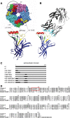
Figure 1.
Figure 1.
Structures of MIR chimeras. A, Putative MIR components are highlighted on the crystal structure of an Aplysia AChBP subunit (Hansen et al., 2005). A top-view space-filling model is shown at the top. A front-view ribbon diagram, rotated to permit viewing of the MIR from its underside as a prominent appendage, is shown at the bottom. Red highlights the α helical ribbon corresponding to α1 1–14. The thin segment proceeding the α helix represents a FLAG tag on the AChBP constructs. Yellow highlights sequence corresponding to α1 15–32. Green highlights sequence corresponding to α1 60–81, including the MIR loop α1 66–76. Dark blue designates the remainder of the α1 subunit. A circle with a radius of 15.5Å and center at the MIR indicates the area typically buried upon antibody binding to a protein antigen (Mariuzza et al., 1987; Konstantakaki et al., 2007). The prominent loop on the right is the C loop which closes over the ACh binding site when agonists are bound. B, The crystal structure of a Fab of mAb 192 (Kontou et al., 2000) is accompanied by the mouse α1 extracellular domain (Dellisanti et al., 2007). This allows comparison between the similar structures of the α1 extracellular domain and AChBP subunit and it allows comparison between the size of an Fab and the MIR. This makes it evident that while very small differences in the sequence and conformation of epitopes can profoundly influence the affinity with which antibodies are bound, the large size of bivalent IgG molecules can result in competitive binding between different closely spaced epitopes within the MIR. Parts of the MIR are highlighted in the same colors as in the AChBP subunit. The light chain of the Fab is gray and the heavy chain is black. Six hypervariable loops, which form the antigen binding sites, are highlighted in cyan. This mAb to the MIR does not bind to the MIR loop but competes for binding with mAbs which do. The Fab is angled to suggest this, but not actually docked on the subunit. Future studies in which the crystal structures of MIR chimeras with Fabs bound are determined should reveal the contact amino acids involved in binding of mAbs to the MIR. C, A schematic diagram of the α1/α7 chimeras used to map MIR epitopes is shown. The extracellular domain of each chimeric subunit is displayed with the α1 sequences indicated by black boxes. D, Alignment of extracellular domain sequences of human and rat muscle AChR α1 subunits. Rodent α1 differs significantly at the sequence α1 23–30 from human α1 (in red), making this sequence a likely candidate for contributing to MIR epitopes which differ between rats and humans, such as the epitope for mAb 192.
Figure 2.
Figure 2.
α1/α7 chimeras were more efficiently expressed than wild-type α7 AChRs. A, Expression on the oocyte surface detected by binding of 125I αBgt to intact oocytes is shown. Presence of the α1 MIR increased assembly of mature α1/α7 AChRs as measured by the total amount of chimeric AChRs on the cell surface. Only mature pentameric AChRs are expected to be transported to the cell surface. All pairs of values shown are significantly different from each other (p < 0.05) except α1(66–76)/α7 and α1(1–32, 60–81)/α7. B, Similar amounts of wild-type α7 and α1/α7 chimera proteins were synthesized as measured by a Western blot (top). However, the α1/α7 chimera which forms the most antigenic MIR structure was assembled much more efficiently into mature AChRs expressed on the oocyte surface (bottom, p < 0.05 for all pairs). A chimera containing only α1 1–32 was not expressed on the cell surface at all. C, The fractions of wild-type α7 and α1(2–14, 60–81)/α7 which could be adsorbed by αBgt-coupled beads were compared on a Western blot using mAb 319 followed by 125I labeled goat anti-rat IgG. Radioactive bands were cut out and quantitated using a γ counter. Only mature pentameric AChRs are capable of binding αBgt. With wild-type α7, 6.6% of subunits were assembled into mature AChRs that were adsorbed to αBgt-coupled beads. Of α1(2–14, 60–81)/α7 subunits, 71% formed mature AChRs. The α1(2–14, 60–81)/α7 chimera always had more protein on Western blots than wild-type α7. One possible explanation is that, as with α1 (Merlie and Lindstrom, 1983), unassembled subunits turn over much more rapidly than mature AChRs. Thus the remarkable assembly efficiency of α1(2–14, 60–81)/α7 (Fig. 2A) results in accumulation of much more total α7 subunits.
Figure 3.
Figure 3.
α1/α7 chimeras greatly influence the sensitivity to activation of chimeric AChRs by ACh. A, Dose/response curves for ACh show that these chimeras exhibited significantly different sensitivities to activation by ACh. Normalized currents are plotted against agonist concentrations. Averaged data (each point from three to five experiments) were fitted by the Hill equation. The single-fit errors were shown. B, The α1 sequences in the chimeras did not change the maximum current per AChR at saturating ACh concentrations of chimeric AChRs. Chimeras were expressed in Xenopus oocytes and function was studied with whole-cell recordings using a standard two-microelectrode voltage clamp. The total amount of chimeric AChRs on the cell surface was detected by binding of 125I αBgt to intact oocytes. C, Time course of currents induced by EC50 concentrations of ACh. Currents were normalized to the peak currents for each chimera.
Figure 4.
Figure 4.
Binding affinity of mAb 637 to the α1(1–30, 60–81)/AChBP chimera is slightly decreased in the presence of saturating nicotine or αBgt. The binding of various concentrations of mAbs to 125I labeled α1/AChBP chimera was determined by immunoprecipitation in the absence (circles) and presence of either 1 mm nicotine (square) or 2.5 μm αBgt (triangle). The averages of duplicate determinations were fitted by the Hill equation to determine KD values (see Table 2).
Figure 5.
Figure 5.
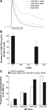
Chimeric α7(66–76)/α1 AChRs lacking the α1 MIR loop were not immunoprecipitable by most mAbs to the MIR or by many MG patient autoantibodies. A, Chimeric α7(66–76)/α1 subunits formed functional AChRs when coexpressed with β1, δ, and either γ or ε subunits. Chimeric α7(66–76)/α1 subunits expressed with γ subunits to produce a fetal type AChR desensitized more rapidly than wild-type fetal AChR. B, 125I αBgt labeled (α7(66–76)/α1)2β1δγ AChRs were immunoprecipitated from Triton X-100 extracts by mAbs 61 and 192, but not by mAbs 198, 210 or 637 (*p < 0.05). mAb 61 is directed at the large cytoplasmic domain of the α1 subunit (Ratnam et al., 1986). C, The chimeric (α7(66–76)/α1)2β1δγ AChR prevented binding of an average of 57% of tested MG sera when compared to wild-type (α1)2β1δγ. It is consistent with inhibition of binding of these sera to AChR by mAb 35 (average 55%). The sera are numbered as in Figure 7B.
Figure 6.
Figure 6.
mAb 637 bound to the MIR on unassembled α1 subunits with low affinity compared with native AChR. The binding of different concentrations of mAb to 125I αBgt labeled Triton X-100 extracts of TE671 cells was determined by immunoprecipitation assay in the presence (open circles) and absence (closed circles) of 10 mm nicotine. The binding curve in the absence of nicotine showed a high and a low affinity component. The low affinity phase did not reach a plateau within the tested concentration range. The monotonic calculated high affinity binding curve, determined by subtracting the low affinity binding in the presence of nicotine, is shown as a dotted line (× symbols). KD1 for high affinity to the MIR in mature AChRs was determined from the monotonic calculated binding curve by fitting with the Hill equation as described in Table 2. KD2 for low affinity binding to unassembled α1 was determined from the binding curve in the presence of nicotine by fitting with a two-component Hill equation (r = (1 − A)/(1 + 10(LogKD1 − Log[ACh]) × Hill slope1) + A/(1 + 10(LogKD2 − Log[ACh]) × Hill slope2), where A is the proportion of the low affinity component). The binding curve in the absence of nicotine was also fit with this two-component Hill equation. Nicotine inhibits binding of 125I αBgt to mature AChRs but not to the unassembled α1 subunits which are present in these extracts. Thus, in the presence of nicotine binding to only unassembled α1 is assayed.
Figure 7.
Figure 7.
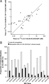
The MIR expressed in chimeric AChBP was recognized by MG patient sera. A, Titers of sera from 61 MG patients were determined by immunoprecipitation of 125I α1(1–30, 60–81)/AChBP or 125I α1(1–30, 60–81, 107–115)/AChBP. A linear regression line was fitted to the data with the following parameters: r = 0.90 and slope = 0.49. These results indicated that both chimeras were recognized equally, implying that the sequence α1 107–115 did not contribute significantly to autoantibody binding. B, The proportion of MG autoantibodies specific to the MIR was determined in sera from 13 other MG patients by two methods. The fraction of autoantibodies to human muscle AChR that could bind to the α1(1–30, 60–81)/AChBP chimera was determined by pre-adsorbing the antisera with the α1(1–30, 60–81)/AChBP chimera coupled to agarose. The fraction of autoantibodies that could be inhibited from binding to AChR by the presence of excess mAb 35 was also determined. Binding of mAb 35 inhibited the binding of a greater proportion of the autoantibodies than could bind the α1 epitopes expressed in the chimera.
Figure 8.
Figure 8.
The MIR in the α1 1–30, 60–81/AChBP chimera was recognized by a majority of feline and canine MG sera. A, C, The apparent titers of 21 canine and 39 feline MG sera against 125I labeled α1(1–30, 60–81)/AChBP were determined by immunoprecipitation assay and reported as nanomoles of 125I labeled α1(1–30, 60–81)/AChBP bound per liter of serum. B, D, Proportion of anti-MIR autoantibodies was expressed as the percentage of each serum titer against 125I labeled α1(1–30, 60–81)/AChBP relative to the corresponding titer against muscle AChR from that species. From left to right, MG sera were arranged in ascending order of titers against muscle AChR.

References

    1. Beroukhim R, Unwin N. Three-dimensional location of the main immunogenic region of the acetylcholine receptor. Neuron. 1995;15:323–331. - PubMed
    1. Castelán F, Mulet J, Aldea M, Sala S, Sala F, Criado M. Cytoplasmic regions adjacent to the M3 and M4 transmembrane segments influence expression and function of α7 nicotinic acetylcholine receptors. A study with single amino acid mutants. J Neurochem. 2007;100:406–415. - PubMed
    1. Castillo M, Mulet J, Aldea M, Gerber S, Sala S, Sala F, Criado M. Role of the N-terminal α-helix in biogenesis of α7 nicotinic receptors. J Neurochem. 2009;108:1399–1409. - PubMed
    1. Colman A. Transcription and translation: a practical approach. In: Hames BD, Higgins SJ, editors. Practical approach series. Oxford, UK: IRL Press; 1984. pp. 271–302.
    1. Conroy WG, Saedi MS, Lindstrom J. TE671 cells express an abundance of a partially mature acetylcholine receptor α subunit which has characteristics of an assembly intermediate. J Biol Chem. 1990;265:21642–21651. - PubMed

Publication types

MeSH terms