

Reduced hippocampal damage and epileptic seizures after status epilepticus in mice lacking proapoptotic Puma
- PMID: 19890018
- PMCID: PMC3231945
- DOI: 10.1096/fj.09-145870
Reduced hippocampal damage and epileptic seizures after status epilepticus in mice lacking proapoptotic Puma
Abstract
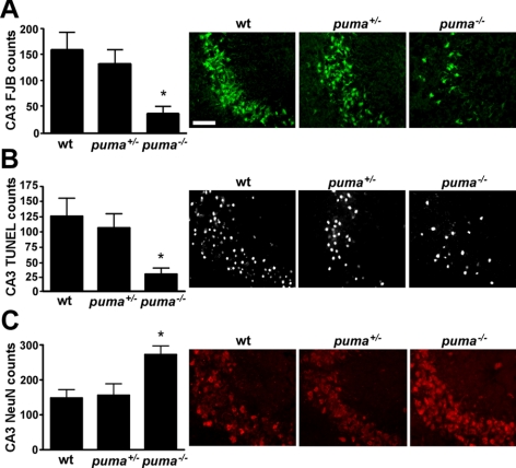
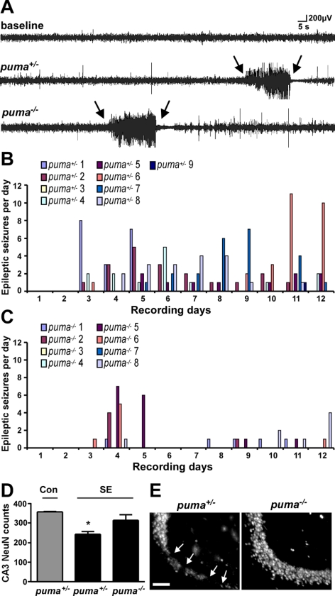
The functional significance of neuronal death for pathogenesis of epilepsy and the underlying molecular mechanisms thereof remain incompletely understood. The p53 transcription factor has been implicated in seizure damage, but its target genes and the influence of cell death under its control on epilepsy development are unknown. In the present study, we report that status epilepticus (SE) triggered by intra-amygdala kainic acid in mice causes rapid p53 accumulation and subsequent hippocampal damage. Expression of p53-up-regulated mediator of apoptosis (Puma), a proapoptotic Bcl-2 homology domain 3-only protein under p53 control, was increased within a few hours of SE. Induction of Puma was blocked by pharmacologic inhibition of p53, and hippocampal damage was also reduced. Puma induction was also blocked in p53-deficient mice subject to SE. Compared to Puma-expressing mice, Puma-deficient mice had significantly smaller hippocampal lesions after SE. Long-term, continuous telemetric EEG monitoring revealed a approximately 60% reduction in the frequency of epileptic seizures in the Puma-deficient mice compared to Puma-expressing mice. These are the first data showing genetic deletion of a proapoptotic protein acting acutely to influence neuronal death subsequently alters the phenotype of epilepsy in the long-term, supporting the concept that apoptotic pathway activation is a trigger of epileptogenesis.-Engel, T., Murphy, B. M., Hatazaki, S., Jimenez-Mateos, E. M., Concannon, C. G., Woods, I., Prehn, J. H. M., Henshall, D. C. Reduced hippocampal damage and epileptic seizures after status epilepticus in mice lacking proapoptotic Puma.
Figures
Similar articles
-
Deletion of Puma protects hippocampal neurons in a model of severe status epilepticus.Neuroscience. 2010 Jun 30;168(2):443-50. doi: 10.1016/j.neuroscience.2010.03.057. Epub 2010 Apr 1. Neuroscience. 2010. PMID: 20362645 Free PMC article.
-
Loss of p53 results in protracted electrographic seizures and development of an aggravated epileptic phenotype following status epilepticus.Cell Death Dis. 2010 Oct 7;1(10):e79. doi: 10.1038/cddis.2010.55. Cell Death Dis. 2010. PMID: 21368852 Free PMC article.
-
Deletion of the BH3-only protein Noxa alters electrographic seizures but does not protect against hippocampal damage after status epilepticus in mice.Cell Death Dis. 2017 Jan 12;8(1):e2556. doi: 10.1038/cddis.2016.301. Cell Death Dis. 2017. PMID: 28079889 Free PMC article.
-
Importance of proapoptotic protein PUMA in cell radioresistance.Folia Biol (Praha). 2014;60(2):53-6. doi: 10.14712/fb2014060020053. Folia Biol (Praha). 2014. PMID: 24785107 Review.
-
PUMA, a potent killer with or without p53.Oncogene. 2008 Dec;27 Suppl 1(Suppl 1):S71-83. doi: 10.1038/onc.2009.45. Oncogene. 2008. PMID: 19641508 Free PMC article. Review.
Cited by
-
Chronic Trigeminal Nerve Stimulation Protects Against Seizures, Cognitive Impairments, Hippocampal Apoptosis, and Inflammatory Responses in Epileptic Rats.J Mol Neurosci. 2016 May;59(1):78-89. doi: 10.1007/s12031-016-0736-5. Epub 2016 Mar 14. J Mol Neurosci. 2016. PMID: 26973056
-
Transgenic overexpression of 14-3-3 zeta protects hippocampus against endoplasmic reticulum stress and status epilepticus in vivo.PLoS One. 2013;8(1):e54491. doi: 10.1371/journal.pone.0054491. Epub 2013 Jan 24. PLoS One. 2013. PMID: 23359526 Free PMC article.
-
TGFβ1 treatment reduces hippocampal damage, spontaneous recurrent seizures, and learning memory deficits in pilocarpine-treated rats.J Mol Neurosci. 2013 May;50(1):109-23. doi: 10.1007/s12031-012-9879-1. Epub 2012 Aug 31. J Mol Neurosci. 2013. PMID: 22936246
-
Bi-directional genetic modulation of GSK-3β exacerbates hippocampal neuropathology in experimental status epilepticus.Cell Death Dis. 2018 Sep 20;9(10):969. doi: 10.1038/s41419-018-0963-5. Cell Death Dis. 2018. PMID: 30237424 Free PMC article.
-
miRNA Expression profile after status epilepticus and hippocampal neuroprotection by targeting miR-132.Am J Pathol. 2011 Nov;179(5):2519-32. doi: 10.1016/j.ajpath.2011.07.036. Epub 2011 Sep 23. Am J Pathol. 2011. PMID: 21945804 Free PMC article.
References
-
- DeGiorgio C. M., Tomiyasu U., Gott P. S., Treiman D. M. Hippocampal pyramidal cell loss in human status epilepticus. Epilepsia. 1992;33:23–27. - PubMed
-
- Fujikawa D. G., Itabashi H. H., Wu A., Shinmei S. S. Status epilepticus-induced neuronal loss in humans without systemic complications or epilepsy. Epilepsia. 2000;41:981–991. - PubMed
-
- Mikaeloff Y., Jambaque I., Hertz-Pannier L., Zamfirescu A., Adamsbaum C., Plouin P., Dulac O., Chiron C. Devastating epileptic encephalopathy in school-aged children (DESC): a pseudo encephalitis. Epilepsy Res. 2006;69:67–79. - PubMed
-
- Pitkanen A., Kharatishvili I., Karhunen H., Lukasiuk K., Immonen R., Nairismagi J., Grohn O., Nissinen J. Epileptogenesis in experimental models. Epilepsia. 2007;48(Suppl. 2):13–20. - PubMed
Publication types
MeSH terms
Substances
Grants and funding
LinkOut - more resources
Full Text Sources
Other Literature Sources
Medical
Research Materials
Miscellaneous
