. 2009 Dec 1;183(11):7169-77.
doi: 10.4049/jimmunol.0901906. Epub 2009 Nov 4.

Th1, Th17, and Th9 effector cells induce experimental autoimmune encephalomyelitis with different pathological phenotypes

Affiliations

Th1, Th17, and Th9 effector cells induce experimental autoimmune encephalomyelitis with different pathological phenotypes

Anneli Jäger et al. J Immunol. .

Abstract

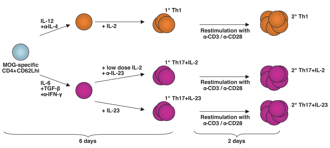
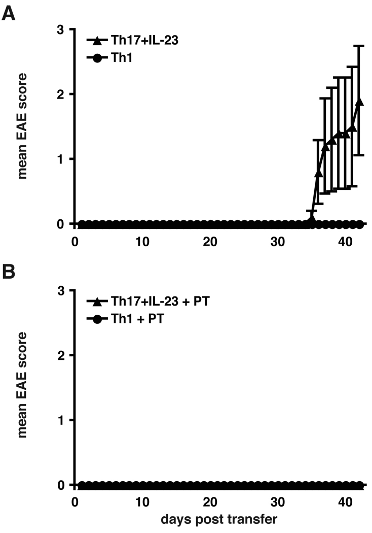
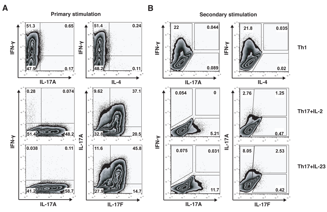
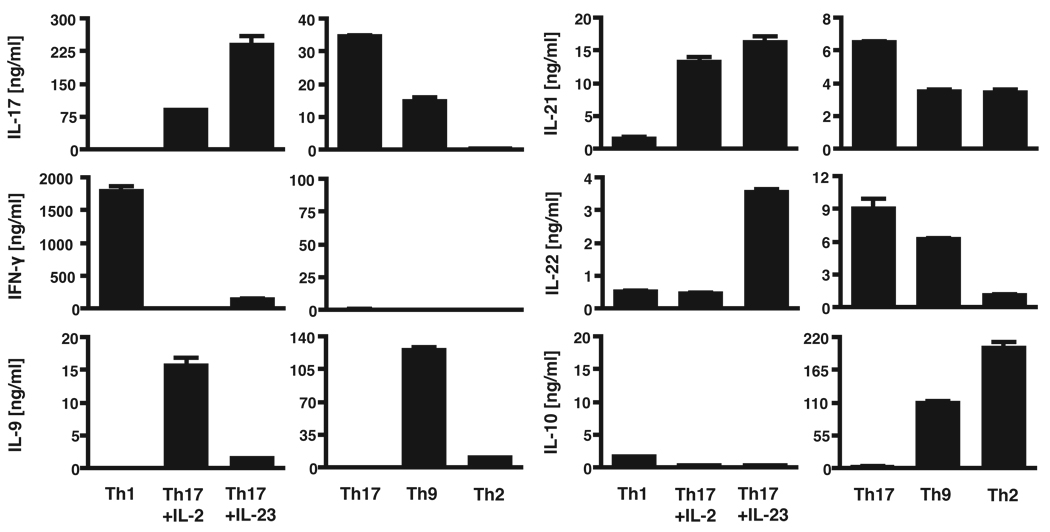
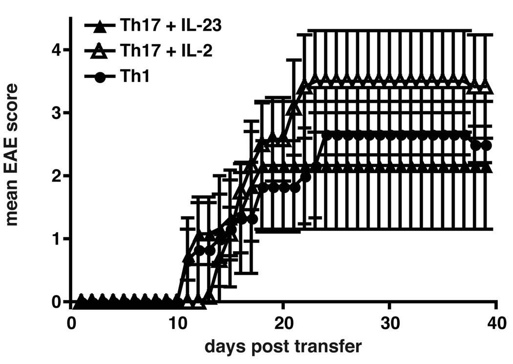
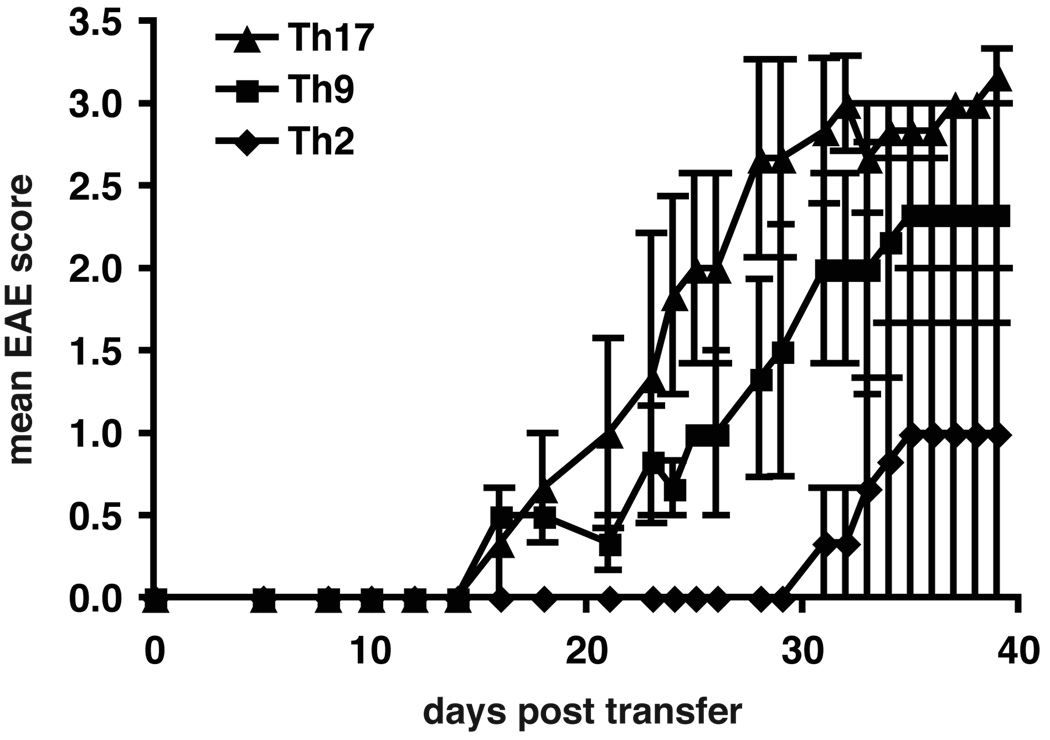
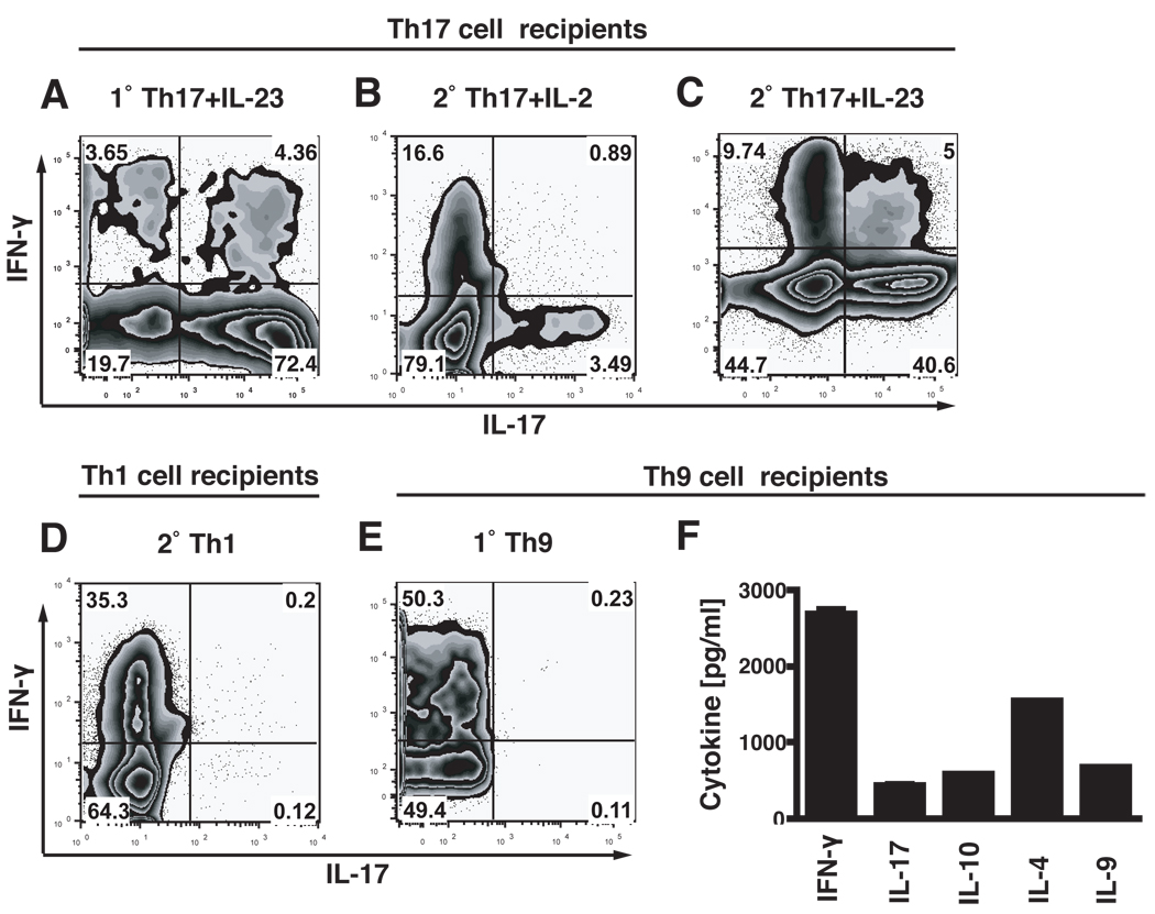
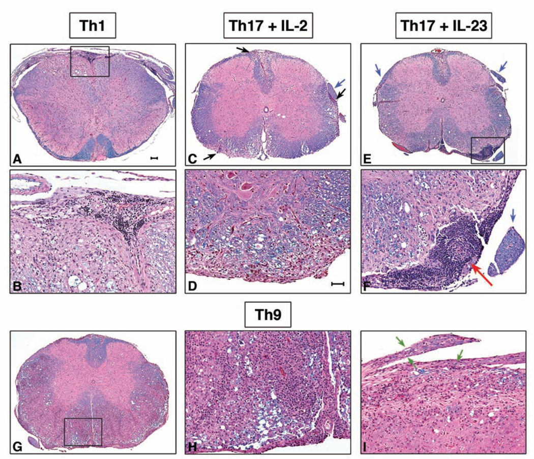
Experimental autoimmune encephalomyelitis (EAE) is a model of human multiple sclerosis induced by autoreactive Th cells that mediate tissue inflammation and demyelination in the CNS. Initially, IFN-gamma-producing Th1 cells and, more recently, IL-17-producing Th17 cells with specificity for myelin Ags have been implicated in EAE induction, but whether Th17 cells are encephalitogenic has been controversial. Moreover, a new effector T cell subset, Th9 cells, has been identified; however, the ability of this T cell subset to induce EAE has not been investigated. Here, we have developed protocols to generate myelin oligodendrocyte glycoprotein-specific Th17, Th1, Th2, and Th9 cells in vitro, so that we could directly compare and characterize the encephalitogenic activity of each of these subsets upon adoptive transfer. We show that myelin oligodendrocyte glycoprotein-specific Th1, Th17, and Th9 cells but not Th2 cells induce EAE upon adoptive transfer. Importantly, each T cell subset induced disease with a different pathological phenotype. These data demonstrate that different effector T cell subsets with specificity for myelin Ags can induce CNS autoimmunity and that the pathological heterogeneity in multiple sclerosis lesions might in part be due to multiple distinct myelin-reactive effector T cells.


Conflict of interest statement

Disclosures

The authors have no financial conflict of interest.

Figures

Figure 1
Figure 1. Generation of T cell subsets in vitro
MOG-specific CD4+CD62Lhi T cells were differentiated into Th1 and Th17 cells. After initial activation in the presence of the indicated polarizing cytokines and neutralizing Abs for 2 days, Th1 cells were further cultured with IL-2, while Th17 cells were further supplemented either with IL-23 or with low doses of IL-2. After 6 days of culture primary (1°) Th1, Th17 plus IL-2, and Th17 plus IL-23 cells were either collected for transfer, or re-stimulated with Abs to CD3 and CD28 for 2 days to generate highly activated secondary (2°) Th1, Th17 plus IL-2, and Th17 plus IL-23 cells.
Figure 2
Figure 2. Transfer of primary Th1 and Th17 cells
A, primary Th17 cells but not Th1 cells can transfer EAE. MOG-specific CD4+CD62Lhi T cells obtained from 2D2 TCR transgenic mice were stimulated with irradiated APCs and anti-CD3 and differentiated into Th1 or Th17 cells with polarizing cytokines. Th17 cells were additionally supplemented with IL-23. After 5 days 4×10⁶ cytokine producing cells were injected i.v. into C57Bl/6 recipients. Recipient animals were observed for the development of clinical signs of EAE for 42 days. Error bars represent SEM. The data are representative of 2 independent experiments. B, recipients of primary Th1 and Th17 cells were administered pertussis toxin at the time of T cell transfer to study the effects of pertussis toxin on the induction of EAE by Th1 versus Th17 cells. Experiments were performed as in A, but recipient mice received 150 ng pertussis toxin i.p. on day 0 and 2 after transfer.
Figure 3
Figure 3. Intracellular cytokine profile of in vitro differentiated T cell subsets
In vitro generated Th1 cells and Th17 cells are highly differentiated and produce only subset-specific cytokines. MOG-specific CD4+CD62Lhi T cells were stimulated with irradiated APCs and anti-CD3 and differentiated into Th1 or Th17 cells with polarizing cytokines. After 2 days Th17 cells were supplemented either with low doses of IL-2 or with IL-23 for additional 4 days. A, in the primary stimulation protocol T cells were analyzed for the production of cytokines by intracellular cytokine staining after 4 days of in vitro differentiation. B, in the secondary stimulation protocol T cells were analyzed for the production of cytokines by intracellular cytokine staining after 2 days of re-stimulation with anti-CD3 and anti-CD28 Abs.
Figure 4
Figure 4. Cytokine profiles of in vitro differentiated T cell subsets
In vitro generated secondary Th1, Th17, Th2 and Th9 cells are highly differentiated and produce subset-specific cytokines. MOG-specific CD4+CD62Lhi T cells were differentiated into Th1, Th17, Th9 and Th2 cells with polarizing cytokines. 48 h after re-stimulation the amounts of IL-17, IFN-γ, IL-9, IL-21, IL-22 and IL-10 secreted into the cell culture medium were determined by ELISA.
Figure 5
Figure 5. Induction of EAE with secondary Th1 and Th17 cells
Secondary Th1, Th17 plus IL-2 and Th17 plus IL-23 cells induce EAE with similar severity and onset upon adoptive transfer. MOG-specific CD4+CD62Lhi T cells were stimulated with irradiated APCs and anti-CD3 and differentiated into Th1 or Th17 cells with polarizing cytokines. Th17 cells were supplemented either with low doses of IL-2 or with IL-23. After 7 days cells were re-stimulated in the presence of anti-CD3 and anti-CD28 Abs for 48h. 3×10⁶ cytokine producing cells were injected i.v. into C57Bl/6 recipients. Recipient animals were observed for the development of clinical signs of EAE for 40 days. Shown are the mean clinical scores of one experiment, error bars represent SEM. Similar results were obtained in 3 independent experiments.
Figure 6
Figure 6. Induction of EAE with secondary Th17, Th9 and Th2 cells
Secondary Th9 and Th17 cells induce EAE upon adoptive transfer, while secondary Th2 cells induce no or mild/delayed disease. MOG-specific CD4+CD62Lhi T cells were stimulated with irradiated APCs and MOG35-55 and differentiated in vitro into Th17, Th9 or Th2 cells with polarizing cytokines. Th17 cells were supplemented with IL-23. Th9 and Th2 cells were supplemented with IL-2. After 6 days cells were re-stimulated with anti-CD3 and anti-CD28 Abs for 48h without cytokines. 5×10⁶ cells were injected i.v. into C57Bl/6 recipients that received pertussis toxin. Recipient animals were observed for the development of clinical signs of EAE for 40 days. Shown are the mean clinical scores of one experiment, error bars represent SEM. Similar results were obtained in 2 independent experiments.
Figure 7
Figure 7. Cytokine profiles of transferred cells recovered from the CNS
At the peak of disease infiltrating cells were isolated from brain and spinal cord of recipient mice. Cells were stimulated with PMA/ionomycin for 4.5 h in the presence of monensin and transferred T cells, identified by their expression of Vα-3.2 TCR, were analyzed for the production of IL-17 and IFN-γ by intracellular cytokine staining. Cells recovered from recipients of 1°Th17 cells cultured with IL-23 produced mostly IL-17 und only little IFN-γ (A). Cells recovered from recipients of 2°Th17 cells cultured with low doses of IL-2 had decreased their IL-17 production and produced IFN-γ (B), whereas cells recovered from 2°Th17 plus IL-23 recipients maintained their IL-17 production stable but also produced some IFN-γ (C). Cells recovered from 2°Th1 cell recipients produced only IFN-γ and no IL-17 (D). Cells recovered from 2°Th9 cell recipients produced IFN-γ as was determined by intracellular cytokine staining (E). However, when infiltrating cells from Th9 cell recipients were stimulated for 12h with PMA/ionomycin besides IFN-γ also IL-17, IL-10, IL-4 and IL-9 were detected in the culture medium with a cytokine bead array (F). All graphs are representative of at least 2 independent experiments.
Figure 8
Figure 8. Histopathology of representative spinal cord sections from Th1, Th17, and Th9 cell recipient mice
A, B: 2°Th1 cell recipients have typical meningeal and parenchymal mononuclear cell infiltrates predominantly in the white matter. Boxed area in A is shown at 20X magnification in B. There are large numbers of small lymphocytes in the subarachnoid space. Parenchymal demyelination is indicated by loss of blue staining associated with vacuolation and foamy macrophages. C, D: 2°Th17 plus IL-2 cell recipient mice show multiple foci of demyelination in white matter tracts (black arrows) and an intact spinal nerve root (blue arrow). D, 20X magnification of a subpial parenchymal lesion. E, F: 2°Th17 plus IL-23 cell recipient mice have demyelinating lesions in the white matter and large accumulations of small lymphocytes in the leptomeninges (boxed area), but intact spinal nerve roots (blue arrows). F, 20X magnification of boxed area in E shows a lymphoid follicle-like area (red arrow) in the leptomeninges. G–I: 2°Th9 cell recipient mice. G, Large areas of the anterior and lateral white matter are diffusely pink indicating loss of myelin. H, 20X magnification of boxed area in G demonstrates diffuse infiltration of sheets of mononuclear cells in the parenchyma and fewer lymphocytes in the subarachnoid space compared to other groups. I, longitudinal section of spinal cord with two nerve roots showing marked diffuse inflammatory cell infiltration and digestion chambers of Cajal indicative of myelin breakdown and Wallerian degeneration (green arrows). Luxol fast blue-hematoxylin and eosin. Bar in A = 100 µm, also for C, E, G; Bar in D = 50 µm, also for B, D, F, H and I.

References

    1. Serafini B, Rosicarelli B, Magliozzi R, Stigliano E, Aloisi F. Detection of ectopic B-cell follicles with germinal centers in the meninges of patients with secondary progressive multiple sclerosis. Brain Pathol. 2004;14:164–174. - PMC - PubMed
    1. Lassmann H, Brück W, Lucchinetti CF. The immunopathology of multiple sclerosis: an overview. Brain Pathol. 2007;17:210–218. - PMC - PubMed
    1. Ben-Nun A, Wekerle H, Cohen IR. The rapid isolation of clonable antigen-specific T lymphocyte lines capable of mediating autoimmune encephalomyelitis. Eur J Immunol. 1981;11:195–199. - PubMed
    1. Merrill JE, Kono DH, Clayton J, Ando DG, Hinton DR, Hofman FM. Inflammatory leukocytes and cytokines in the peptide-induced disease of experimental allergic encephalomyelitis in SJL and B10.PL mice. Proc Natl Acad Sci U S A. 1992;89:574–578. - PMC - PubMed
    1. Ando DG, Clayton J, Kono D, Urban JL, Sercarz EE. Encephalitogenic T cells in the B10.PL model of experimental allergic encephalomyelitis (EAE) are of the Th-1 lymphokine subtype. Cell Immunol. 1989;124:132–143. - PubMed

Publication types

MeSH terms