Review
. 2009 Aug;14(8 Suppl 7):8-13; discussion 16-8.
doi: 10.1017/s1092852900024901.

Role of mitochondria in neurodegenerative diseases: mitochondria as a therapeutic target in Alzheimer's disease

Affiliations
Review

Role of mitochondria in neurodegenerative diseases: mitochondria as a therapeutic target in Alzheimer's disease

P Hemachandra Reddy. CNS Spectr. 2009 Aug.

Abstract

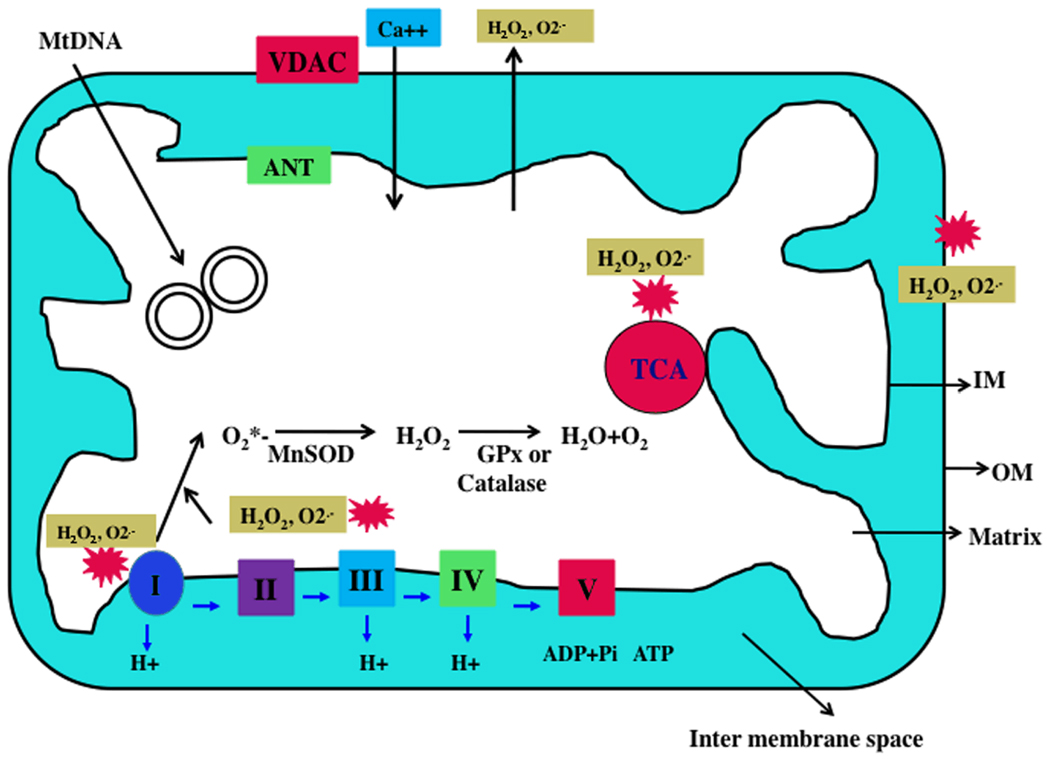
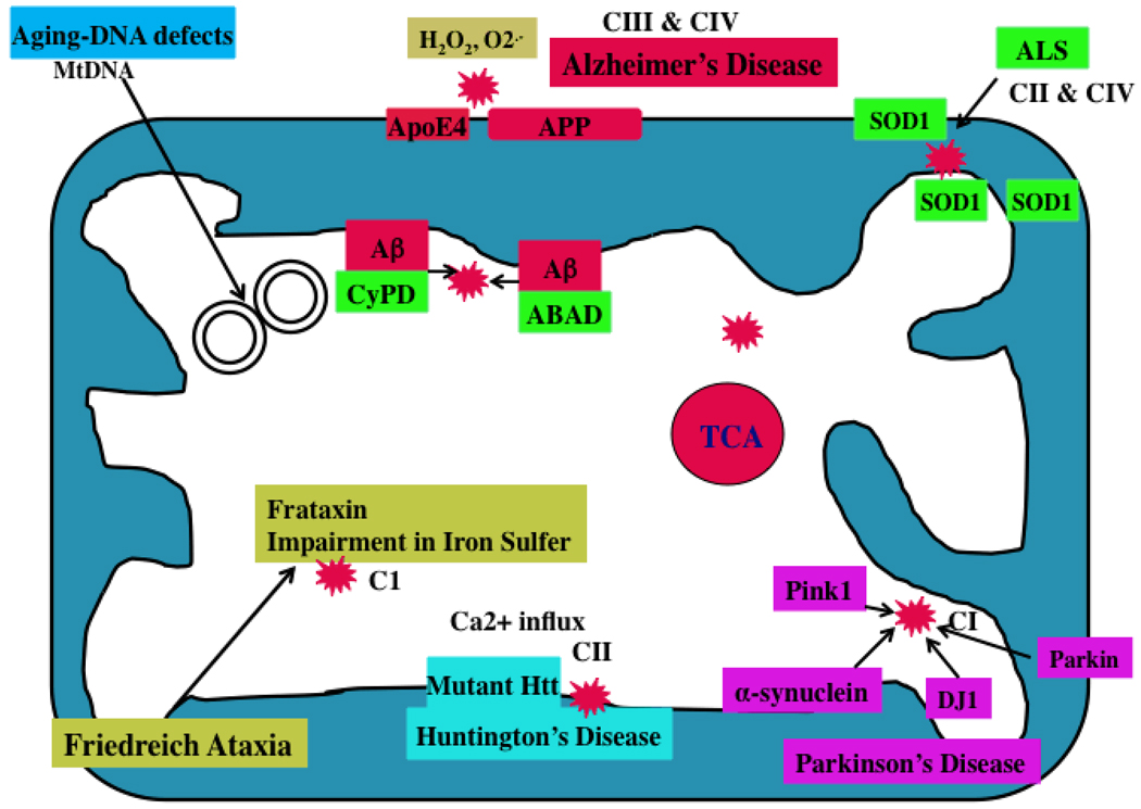
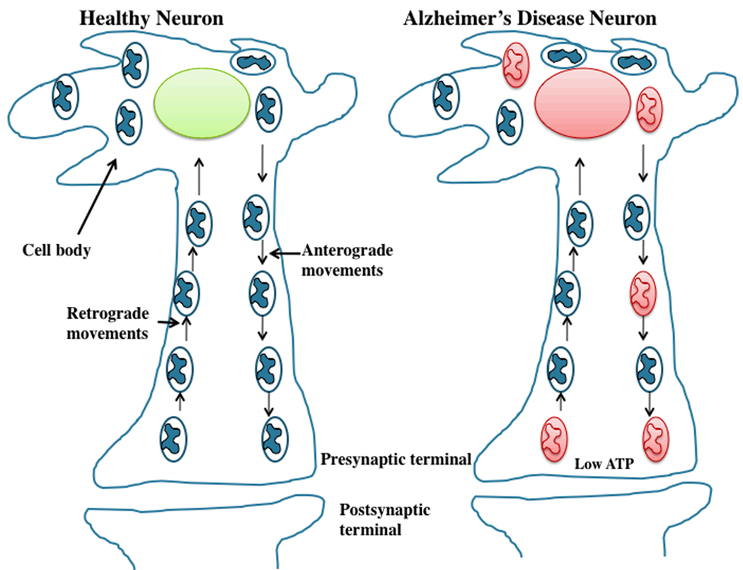
A growing body of evidence suggests that mitochondrial abnormalities are involved in aging and in age-related neurodegenerative diseases as well as cancer, diabetes, and several other diseases known to be affected by mitochondria. Causal factors for most age-related neurodegenerative diseases-including Alzheimer's disease, Parkinson's disease, amyotrophic lateral sclerosis (ALS), and Friedrich ataxia (FRDA)-are largely unknown. Genetic defects are reported to cause a small number of neurodegenerative diseases, but cellular, molecular, and pathological mechanisms of disease progression and selective neuronal cell death are not understood fully in these diseases. However, based on several cellular, molecular, and animal model studies of Alzheimer's disease, Parkinson's disease, ALS, FRDA, cancer, and diabetes, aging may play a large role in cell death in these diseases. Age-dependent, mitochondrially-generated reactive oxygen species (ROS) have been identified as important factors responsible for disease progression and cell death, particularly in late-onset diseases, in which genetic mutations are not causal factors.


Figures

Figure 1
Figure 1. Structure of mitochondria
A mitochondrion is compartmentalized with two lipid membranes: the inner mitochondrial membrane and the outer mitochondrial membrane. The inner mitochondrial membrane houses the mitochondrial respiratory chain and provides a highly efficient barrier to ionic flow. In the ETC, complexes I and III leak electrons to oxygen, producing primarily superoxide radicals. Superoxide radicals are dismutated by manganese superoxide dismuase and produce H2O2. In addition, ETC involves H2O2 reducing to H2O and O2 by catalase or glutathione peroxidase accepting electrons donated by NADH and FADH2 and then yielding energy to generate ATP from adenosine diphosphate and inorganic phosphate. Free radicals are also generated by tricarboxylic acid in the matrix.
Figure 2
Figure 2. Mutant proteins and mitochondria
In AD, Aβ peptides enter mitochondria and interact with mitochondrial proteins, induce free radicals, decrease cytochrome oxidase activity, and inhibit ATP generation. In AD brains, APP is transported to outer mitochondrial membranes, blocks the import of nuclear cytochrome oxidase proteins to mitochondria, and may be responsible for decreased cytochrome oxidase activity. In HD neurons, mutant Htt binds to the outer mitochondrial membrane and induces free radical production. Free radicals may interrupt with calcium uptake. In PD neurons, mutant proteins of α-synuclein, parkin, PINK1, and DJ1 are associated with mitochondria and cause mitochondrial dysfunction. Complex I activity is inhibited in PD neurons. In ALS, mutant SOD1 is localized to the inner and outer mitochondrial membranes and matrix, and induces free radical production and oxidative damage. Impairment of complexes II and IV are associated with ALS. Frataxin is a mitochondrial protein responsible for heme biosynthesis and the formation of iron-sulfur clusters. In Friedriech ataxia, mutant frataxin facilitates the accumulation of iron in mitochondria and induces free radicals.
Figure 3
Figure 3. Abnormal mitochondrial movements in AD neurons
In normal, healthy neurons, mitochondria move from cell body to axons, dendrites, and synapses by an anterograde mechanism, supplying ATP at nerve terminals. Mitochondria then travel back to the cell body from synapses through a retrograde mechanism. In AD neurons, these mechanisms are abnormal primarily due to defective or functionally inactive mitochondria. Mitochondria associated with mutant APP and Aβ are largely dysfunctional and not able to move from body to synapses and supply ATP to nerve terminals (shown in red color). Decreased ATP levels at synapses contribute to synaptic degeneration, synaptic loss and ultimate neuronal death and cognitive impairments.

References

    1. Anandatheerthavarada HK, Biswas G, Robin MA, Avadhani NG. Mitochondrial targeting and a novel transmembrane arrest of Alzheimer's amyloid precursor protein impairs mitochondrial function in neuronal cells. J Cell Biol. 2003;161:41–54. - PMC - PubMed
    1. Anderson S, Bankier AT, Barrell BG, de Bruijn MH, Coulson AR, Drouin J, Eperon IC, Nierlich DP, Roe BA, Sanger F, Schreier PH, Smith AJ, Staden R, Young IG. Sequence and organization of the human mitochondrial genome. Nature. 1981;290:457–465. - PubMed
    1. Beal MF. Mitochondria take center stage in aging and neurodegeneration. Ann Neurol. 2005;58:495–505. - PubMed
    1. Caspersen C, Wang N, Yao J, Sosunov A, Chen X, Lustbader JW, Xu HW, Stern D, McKhann G, Yan SD. Mitochondrial Abeta: a potential focal point for neuronal metabolic dysfunction in Alzheimer's disease. FASEB J. 2005;19:2040–2041. - PubMed
    1. Chandrasekaran K, Giordano T, Brady DR, Stoll J, Martin LJ, Rapoport SI. Impairment in mitochondrial cytochrome oxidase gene expression in Alzheimer disease. Brain Res Mol Brain Res. 1994;24:336–340. - PubMed

Publication types