. 2010 Mar;130(3):682-93.
doi: 10.1038/jid.2009.336. Epub 2009 Nov 5.

EGFR regulates the expression of keratinocyte-derived granulocyte/macrophage colony-stimulating factor in vitro and in vivo

Affiliations

EGFR regulates the expression of keratinocyte-derived granulocyte/macrophage colony-stimulating factor in vitro and in vivo

Francesca Mascia et al. J Invest Dermatol. 2010 Mar.

Abstract

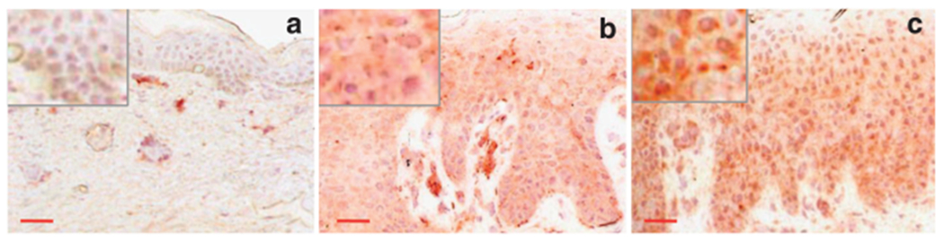
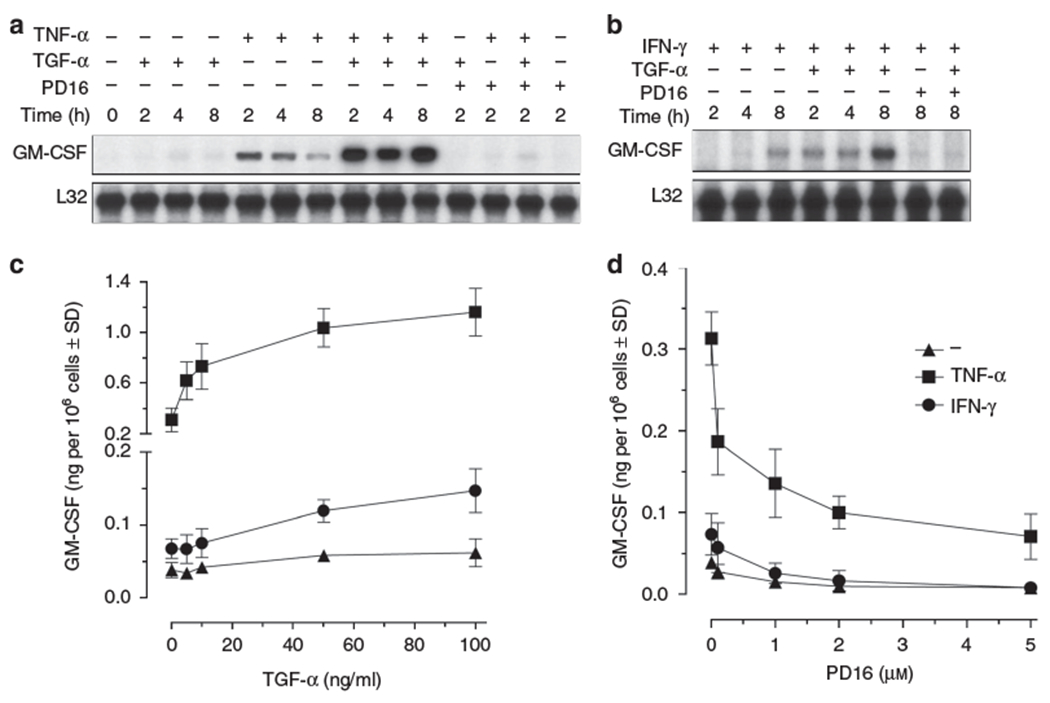
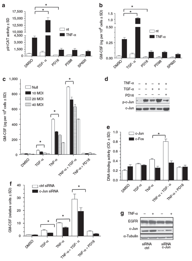
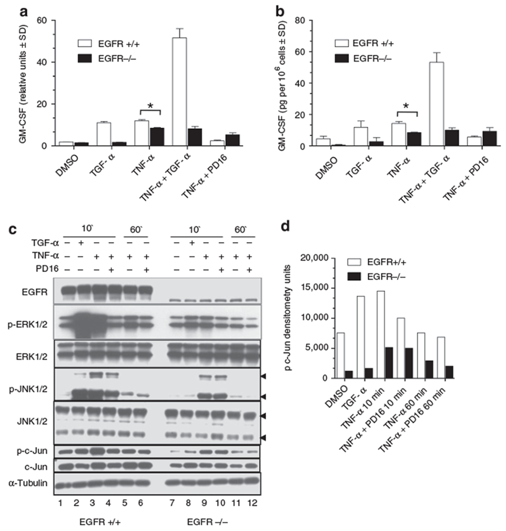
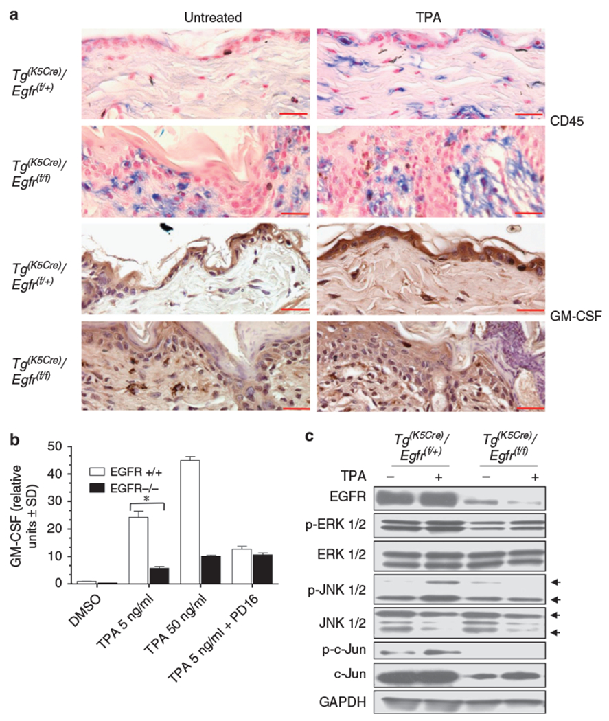
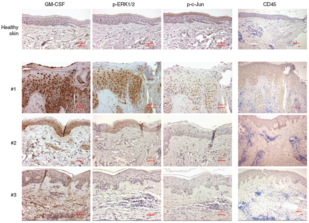
Recent advances in the knowledge of the EGFR pathway have revealed its contribution to distinct immune/inflammatory functions of the epidermis. The purpose of our study was to evaluate the role of EGFR in the regulation of keratinocyte GM-CSF expression. In cultured human keratinocytes, proinflammatory cytokines synergized with TGF-alpha to induce GM-CSF expression. Accordingly, high epidermal levels of EGFR activation are associated with enhanced expression of GM-CSF in lesional skin of patients with psoriasis or allergic contact dermatitis. In cultured keratinocytes, pharmacological inhibition of EGFR activity reduced GM-CSF promoter transactivation, whereas genetic inhibition of AP-1 reduced expression of GM-CSF. Furthermore, EGFR activation enhanced TNF-alpha-induced c-Jun phosphorylation and DNA binding, whereas c-Jun silencing reduced GM-CSF expression. Using two different mouse models, we showed that the lack of a functional EGFR pathway was associated with reduced cytokine-induced phosphorylation of ERK1/2, JNK1/2, c-Jun and reduced keratinocyte-derived GM-CSF expression both in vitro and in vivo. Finally, the analysis of GM-CSF expression in the skin of cancer patients treated with anti EGFR drugs showed an association between ERK activity, c-Jun phosphorylation, and epidermal GM-CSF expression. These data demonstrate that the EGFR pathway is critical for the upregulation of keratinocyte GM-CSF expression under conditions of cytokine stimulation.


Conflict of interest statement

CONFLICT OF INTEREST

The author states no conflict of interest.

Figures

Figure 1.
Figure 1.. GM-CSF is highly expressed by epidermal keratinocytes in chronically inflamed skin.
Expression of GM-CSF in the epidermis of a healthy donor (a), in lesional skin of plaque psoriasis (b) and chronic allergic contact dermatitis (c). Representative immunohistochemistry results from seven healthy donors, five patients with psoriasis, and three patients with allergic contact dermatitis. Normalization bar = 100 μm for panel a (× 200) and 50 μm for panels b and c (× 100).
Figure 2.
Figure 2.. TGF-α enhances, whereas EGFR signaling inhibition suppresses, the expression of GM-CSF by cytokine-stimulated human keratinocytes.
Cytokine gene expression was measured by RNase protection assay in total RNA (10 μg) extracted from human keratinocytes stimulated with TNF-α (50 ng/ml) (a) or IFN-γ (50 U/ml) (b) and/or TGF-α (50 ng/ml), directly or following 30 minutes incubation with PD168393 (1 μm). The autoradiographies are representative of four independent experiments. ELISA detection of GM-CSF levels after 24 hours stimulation of human keratinocytes with escalating doses of TGF-α (c) or PD168393 (PD16) (d). Results are expressed as the mean of three independent experiments ± SD.
Figure 3.
Figure 3.. TGF-α upregulates GM-CSF transcription via increased c-Jun phosphorylation and transactivation.
(a) CAT activity was determined in human keratinocytes transfected with the minimal GM-CSF promoter p91CAT following 6 hours stimulation with 50 ng/ml TNF-α and/or 50 ng/ml TGF-α directly, or following 30 minutes pre-incubation with 2 μ/M PD168393 (PD16), or 20 μm PD98059 (PD98) or SP600125 (SP600). (b) ELISA of GM-CSF levels in the supernatant of keratinocytes after 24 hours stimulation with same conditions as panel a. (c) GM-CSF ELISA detection of culture supernatants 48 hours after infection of human keratinocytes with increasing doses of A-FOS or null adenovirus. Cells were treated in the last 24 hours with TNF-α (50 ng/ml) and/or TGF-α (50 ng/ml), directly or following 30 minutes incubation with PD168393 (1 μm). (d) Twenty μg of total cell lysates were run in an immunoblot to detect phospho c-Jun (ser 63) levels or used to evaluate the relative DNA binding activity versus an AP-1 binding site in (e) where c-Jun/c-Fos DNA binding was tested by TransAM assay. For these experiments, keratinocytes were treated for 15 minutes (panel d) or 30minutes (panel e) with TNF-α (50 ng/ml) and/or TGF-α (50 ng/ml), directly or following 30 minutes incubation with PD168393 (1 μm). (f) GM-CSF mRNA levels were quantified by real-time RT-PCR with and without c-Jun silencing in human keratinocytes stimulated for 4 hours with TNF-α (50 ng/ml) and/or TGF-α (50 ng/ml), directly or following 30 minutes incubation with PD168393 (1 μm). (g) Total levels of c-Jun and EGFR were detected via immunoblot as control for silencing experiment. Values in (a-c) and (f) are expressed as the mean of experimental duplicates± SD. *P<0.01. Panels d and g show one representative experiment. All experiments were repeated at least three times.
Figure 4.
Figure 4.. Genetic ablation of EGFR impairs TNF-α-induced GM-CSF expression and ERK1/2, JNK1/2, and c-Jun phosphorylation in murine keratinocytes.
(a) real-time RT-PCR for murine GM-CSF on wild-type and EGFR null keratinocytes. Cells were stimulated for 1 hour with TNF-α (50 ng/ml) and/or TGF-α (50 ng/ml), directly or following 30 minutes incubation with PD168393 (1 μm). (b) ELISA of cell supernatant after 24 hours stimulation as panel a. (c) Keratinocytes from wild-type (lanes 1–6) and EGFR null mice (lanes 7–12) were stimulated for 10 minutes and 1 hour with TNF-α (50 ng/ml) or TGF-α (50 ng/ml), directly or following 30 minutes incubation with PD168393 (1 μm). Twenty μg of cell lysates were subjected to immunoblotting. (d) Densitometric analysis of phospho c-Jun bands normalized over total c-Jun bands of immunoblot from panel c. Values in (a) and (b) are expressed as the mean of experimental duplicates ± SD. *P<0.01. Experiments were repeated at least three times with one representative experiment shown in the figure.
Figure 5.
Figure 5.. Skin-targeted deletion of EGFR impairs TPA-induced epithelial GM-CSF expression in vivo.
(a) Immunohistochemical detection of CD45 in skin sections from double transgenic wild-type (Tg(K5Cre)/Egft(f/+)) and double transgenic EGFR deleted (Tg(K5Cre)/Egfr(f/f)) mice, untreated and treated with 10 μg of TPA for 7 hours. Immunohistochemical detection of GM-CSF in skin sections from double transgenic wild-type and double transgenic EGFR deleted mice, untreated and treated with 10 μg of TPA for 7 hours. Normalization bar = 50 μm. (b) real-time RT-PCR for murine GM-CSF on wild-type and EGFR null keratinocytes. Cells were stimulated for 1 hour with 5 and 50 ng/ml of TPA directly or following 30 minutes incubation with PD168393 1 μm. (c) Double transgenic wild-type (Tg(K5Cre)/Egft(f/+)) and double transgenic EGFR deleted (Tg(K5Cre)/Egfr(f/f)) mice were treated for 2 hours with vehicle alone or 10 μg of TPA and 50 μg of tissue lysates were immunoblotted. Experiments were repeated at least three times with one representative experiment showed in the figure. Values in (b) are expressed as the mean of experimental duplicates ± SD. *P<0.01.
Figure 6.
Figure 6.. Epidermal GM-CSF expression levels in the inflamed skin of cetuximab treated patients are associated with phosphorylation levels of ERK1/2 and c-Jun and not with CD45 staining.
Skin sections of three representative patients and one control are shown. Patients were affected by cetuximab-induced skin rash (grade II). GM-CSF, phospho ERK1/2, phospho c-Jun, and CD45 immunohistochemistry were performed on serial sections from the same patient. Normalization bar = 50 μm for × 200 magnification of GM-CSF, phospho ERK1/2, and phospho c-Jun staining. Normalization bar = 25 μm for × 100 magnification of CD45 staining.

Similar articles

Cited by

References

    1. Abdul Rhaman JA, Moodley YP, Phillips MJ (2004) Pulmonary alveolar proteinosis associated with psoriasis and complicated by mycobacterial infection: successful treatment with granulocyte-macrophage colony stimulating factor after a partial response to whole lavage. Respirology 9:419–22 - PubMed
    1. Bonifati C, Carducci M, Cordiali Fei P, Trento E, Sacerdoti G, Fazio M et al. (1994) Correlated increases of TNF-α, IL-6 and GM-CSF levels in suction blister fluids and sera of psoriatic patients-relation with disease severity. Clin Exp Dermatol 19:383–7 - PubMed
    1. Braunstein S, Kaplan G, Gottlieb AB, Schwartz M, Walsh G, Abalos RM et al. (1994) GM-CSF activates regenerative epidermal growth and stimulates proliferation in human skin in vivo. J Invest Dermatol 103:601–4 - PubMed
    1. Breuhahn K, Mann A, Müller G, Wilhelmi A, Schirmacher P, Enk A et al. (2000) Epidermal overexpression of granulocyte-macrophage colony-stimulating factor induces keratinocyte proliferation and apoptosis. Cell Growth Differ 11:111–21 - PubMed
    1. Cianfarani F, Tommasi R, Failla CM, Viviano MT, Annessi G, Papi M et al. (2006) Granulocyte/macrophage colony-stimulating factor treatment of human chronic ulcers promotes angiogenesis associated with de novo vascular endothelial growth factor transcription in the ulcer bed. Br J Dermatol 154:34–41 - PubMed

Publication types

MeSH terms