

The effect of skeletal maturity on the regenerative function of intrinsic ACL cells
- PMID: 19890988
- PMCID: PMC2845722
- DOI: 10.1002/jor.21018
The effect of skeletal maturity on the regenerative function of intrinsic ACL cells
Abstract
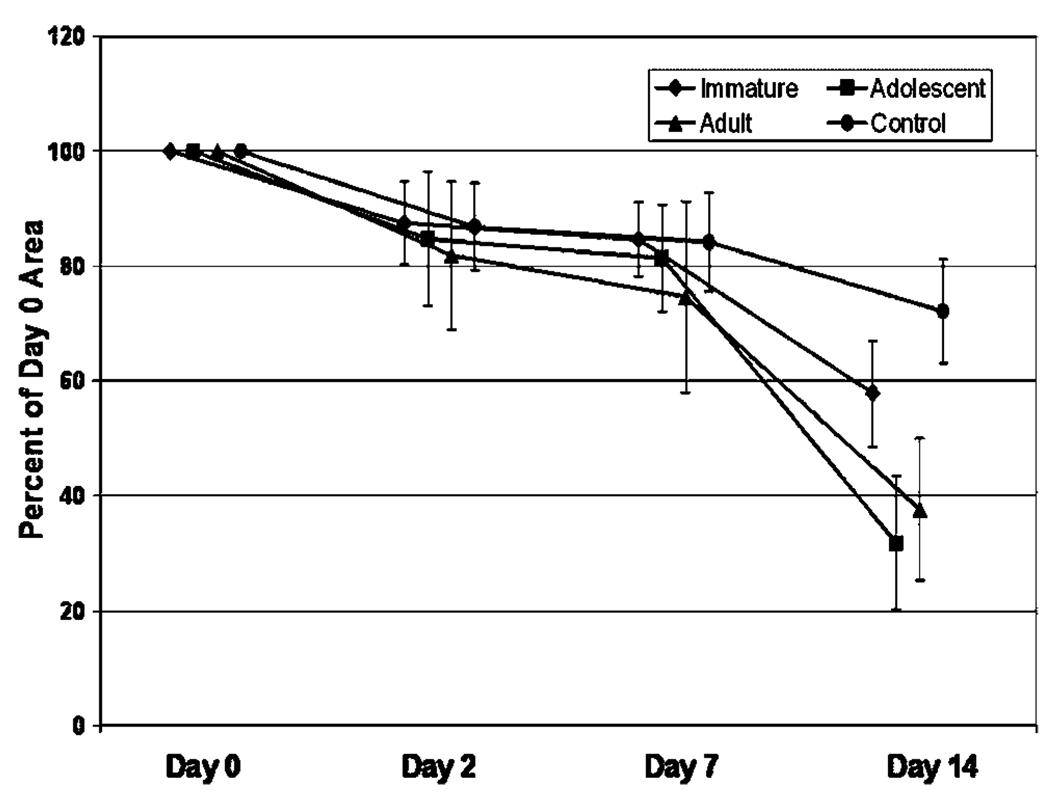
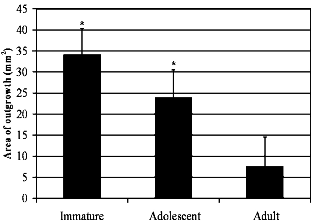
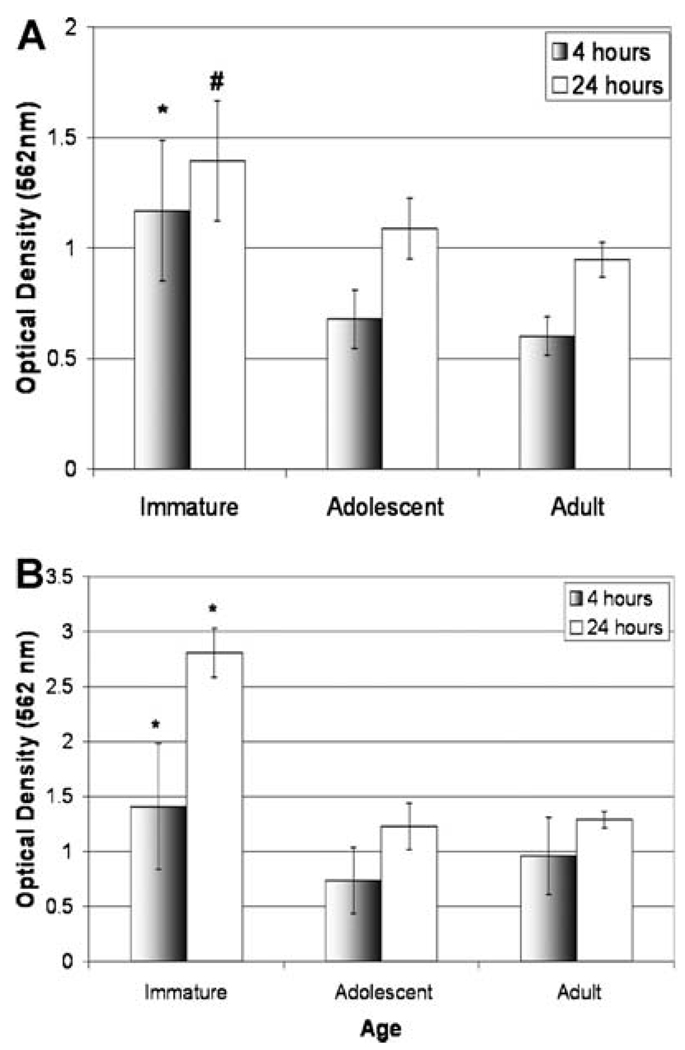
Anterior cruciate ligament (ACL) injuries are an important clinical problem, particularly for adolescent patients. The effect of skeletal maturity on the potential for ACL healing is as yet unknown. In this study, we hypothesized that fibroblastic cells from the ACLs of skeletally immature animals would proliferate and migrate more quickly than cells from adolescent and adult animals. ACL tissue from skeletally immature, adolescent, and adult pigs and sheep were obtained and cells obtained using explant culture. Cell proliferation within a collagen-platelet scaffold was measured at days 2, 7, and 14 of culture using AM MTT assay. Cellular migration was measured at 4 and 24 h using a modified Boyden chamber assay, and cell outgrowth from the explants also measured at 1 week. ACL cells from skeletally immature animals had higher proliferation between 7 and 14 days (p<0.01 for all comparisons) and higher migration potential at all time points in both species (p<0.01 for all comparisons). ACL cells from skeletally immature animals have greater cellular proliferation and migration potential than cells from adolescent or adult animals. These experiments suggest that skeletal maturity may influence the biologic repair capacity of intrinsic ACL cells.
Copyright (c) 2009 Orthopaedic Research Society.
Figures

Similar articles
-
The migration of cells from the ruptured human anterior cruciate ligament into collagen-glycosaminoglycan regeneration templates in vitro.Biomaterials. 2001 Sep;22(17):2393-402. doi: 10.1016/s0142-9612(00)00426-9. Biomaterials. 2001. PMID: 11511036
-
Migration of cells from human anterior cruciate ligament explants into collagen-glycosaminoglycan scaffolds.J Orthop Res. 2000 Jul;18(4):557-64. doi: 10.1002/jor.1100180407. J Orthop Res. 2000. PMID: 11052491
-
Immature animals have higher cellular density in the healing anterior cruciate ligament than adolescent or adult animals.J Orthop Res. 2010 Aug;28(8):1100-6. doi: 10.1002/jor.21070. J Orthop Res. 2010. PMID: 20127960 Free PMC article.
-
The potential for primary repair of the ACL.Sports Med Arthrosc Rev. 2011 Mar;19(1):44-9. doi: 10.1097/JSA.0b013e3182095e5d. Sports Med Arthrosc Rev. 2011. PMID: 21293237 Free PMC article. Review.
-
Anterior cruciate ligament injuries in the skeletally immature patient.Instr Course Lect. 1998;47:351-9. Instr Course Lect. 1998. PMID: 9571437 Review.
Cited by
-
Sex Influences the Biomechanical Outcomes of Anterior Cruciate Ligament Reconstruction in a Preclinical Large Animal Model.Am J Sports Med. 2015 Jul;43(7):1623-31. doi: 10.1177/0363546515582024. Epub 2015 May 4. Am J Sports Med. 2015. PMID: 25939612 Free PMC article.
-
Age dependence of expression of growth factor receptors in porcine ACL fibroblasts.J Orthop Res. 2010 Aug;28(8):1107-12. doi: 10.1002/jor.21111. J Orthop Res. 2010. PMID: 20186834 Free PMC article.
-
Effects of age and platelet-rich plasma on ACL cell viability and collagen gene expression.J Orthop Res. 2012 Jan;30(1):79-85. doi: 10.1002/jor.21496. Epub 2011 Jul 11. J Orthop Res. 2012. PMID: 21748791 Free PMC article.
-
Validation of porcine knee as a sex-specific model to study human anterior cruciate ligament disorders.Clin Orthop Relat Res. 2015 Feb;473(2):639-50. doi: 10.1007/s11999-014-3974-2. Epub 2014 Oct 1. Clin Orthop Relat Res. 2015. PMID: 25269532 Free PMC article.
-
Investigating Commercial Filaments for 3D Printing of Stiff and Elastic Constructs with Ligament-Like Mechanics.Micromachines (Basel). 2020 Sep 11;11(9):846. doi: 10.3390/mi11090846. Micromachines (Basel). 2020. PMID: 32933035 Free PMC article.
References
-
- McIntosh AL, Dahm DL, Stuart MJ. Anterior cruciate ligament reconstruction in the skeletally immature patient. Arthroscopy. 2006;22:1325–1330. - PubMed
-
- Johnston DR, Baker A, Rose C, et al. Long-term outcome of MacIntosh reconstruction of chronic anterior cruciate ligament insufficiency using fascia lata. J Orthop Sci. 2003;8:789–795. - PubMed
-
- Lohmander LS, Ostenberg A, Englund M, et al. High prevalence of knee osteoarthritis, pain, and functional limitations in female soccer players twelve years after anterior cruciate ligament injury. Arthritis Rheum. 2004;50:3145–3152. - PubMed
-
- Aichroth PM, Patel DV, Zorrilla P. The natural history and treatment of rupture of the anterior cruciate ligament in children and adolescents. A prospective review. J Bone Joint Surg Br. 2002;84:38–341. - PubMed
Publication types
MeSH terms
Substances
Grants and funding
LinkOut - more resources
Full Text Sources
Medical
