

Ectopic papilla of Vater in the pylorus
- PMID: 19891024
- PMCID: PMC2773904
- DOI: 10.3748/wjg.15.5221
Ectopic papilla of Vater in the pylorus
Abstract
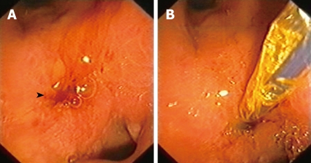
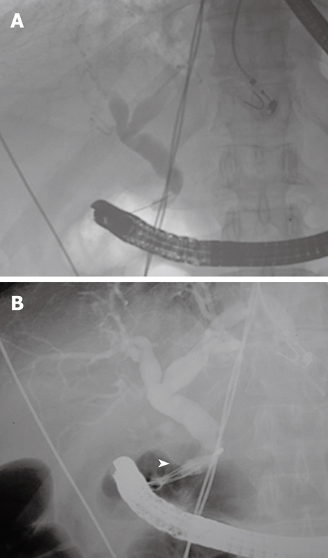
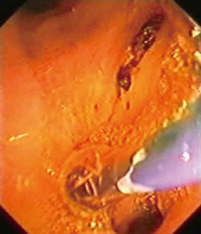
The major papilla of Vater is usually located in the second portion of the duodenum, to the posterior medial wall. Sometimes the mouth of the biliary duct is located in other areas. Drainage of the common bile duct into the pylorus is extremely rare. A 73-year old man, with a history of duodenal ulcer, was admitted to hospital with the diagnosis of cholangitis. Dilatation of the extrahepatic biliary duct was observed by abdominal ultrasonography, and endoscopic retrograde cholangiopancreatography (ERCP) was performed. No area suggesting the presence of the papilla of Vater was found within the second duodenal portion. Finally the major papilla was located in the theoretical pyloric duct. Cholangiography was performed and choledocholithiasis was found in the biliary tree. The patient underwent dilatation of the papilla with a balloon tyre and removal of a 7 mm stone using a Dormia basket, which solved the problem without further complications. This anomaly increased the difficulty of performing therapeutic interventions during ERCP. This alteration in anatomy may increase the risk of complications during papillotomy, with a theoretically higher risk of perforation. Dilatation using a balloon was the chosen therapeutic technique both in our case and in the literature, due to its low rate of complications.
2009 The WJG Press and Baishideng. All rights reserved.
Figures
References
-
- Doty J, Hassall E, Fonkalsrud EW. Anomalous drainage of the common bile duct into the fourth portion of the duodenum. Clinical sequelae. Arch Surg. 1985;120:1077–1079. - PubMed
-
- Lee HJ, Ha HK, Kim MH, Jeong YK, Kim PN, Lee MG, Kim JS, Suh DJ, Lee SG, Min YI, et al. ERCP and CT findings of ectopic drainage of the common bile duct into the duodenal bulb. AJR Am J Roentgenol. 1997;169:517–520. - PubMed
Publication types
MeSH terms
LinkOut - more resources
Full Text Sources
Medical
