. 2009 Dec;31(12):741-9.
doi: 10.1111/j.1365-3024.2009.01151.x.

IL-4(-/-) mice with lethal Mesocestoides corti infections--reduced Th2 cytokines and alternatively activated macrophages

Affiliations

IL-4(-/-) mice with lethal Mesocestoides corti infections--reduced Th2 cytokines and alternatively activated macrophages

A E O'Connell et al. Parasite Immunol. 2009 Dec.

Abstract

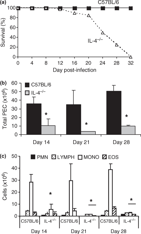
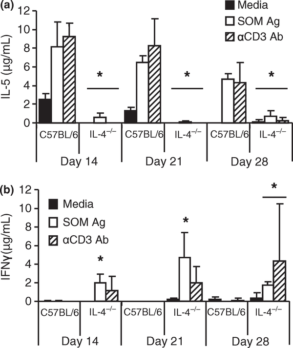
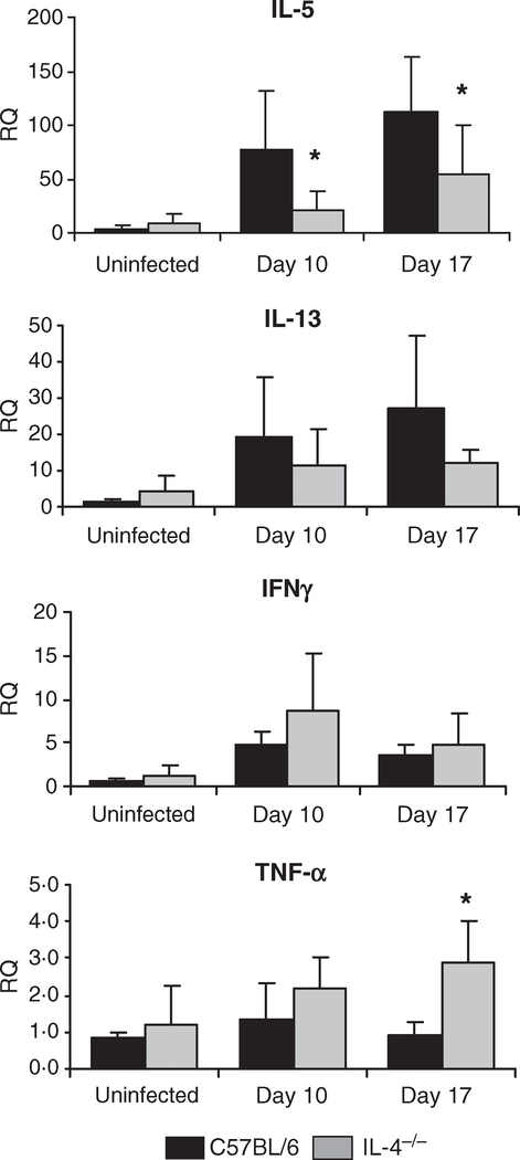
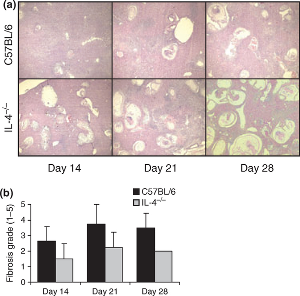
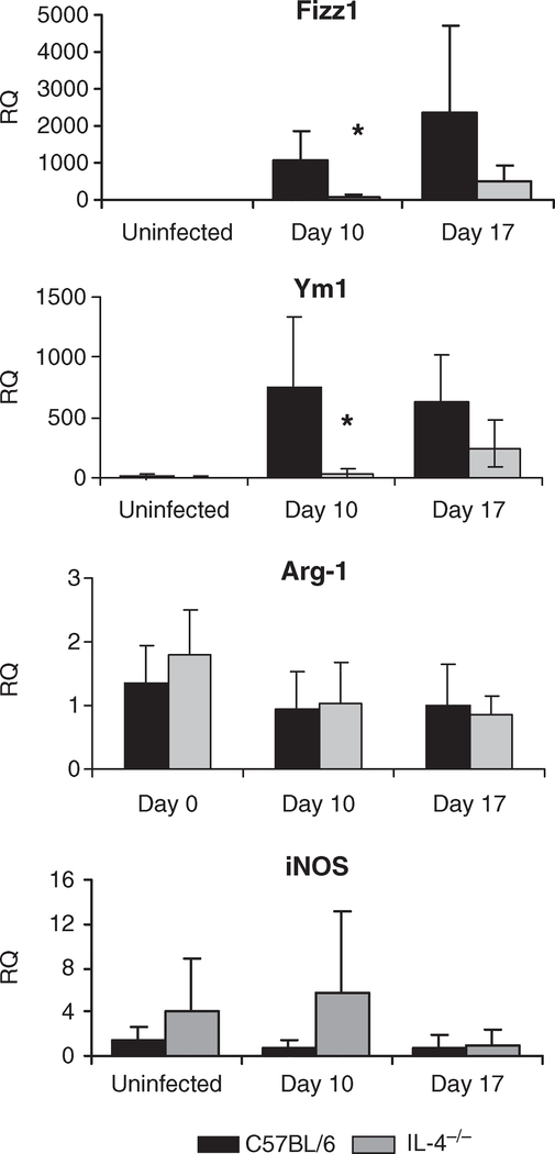
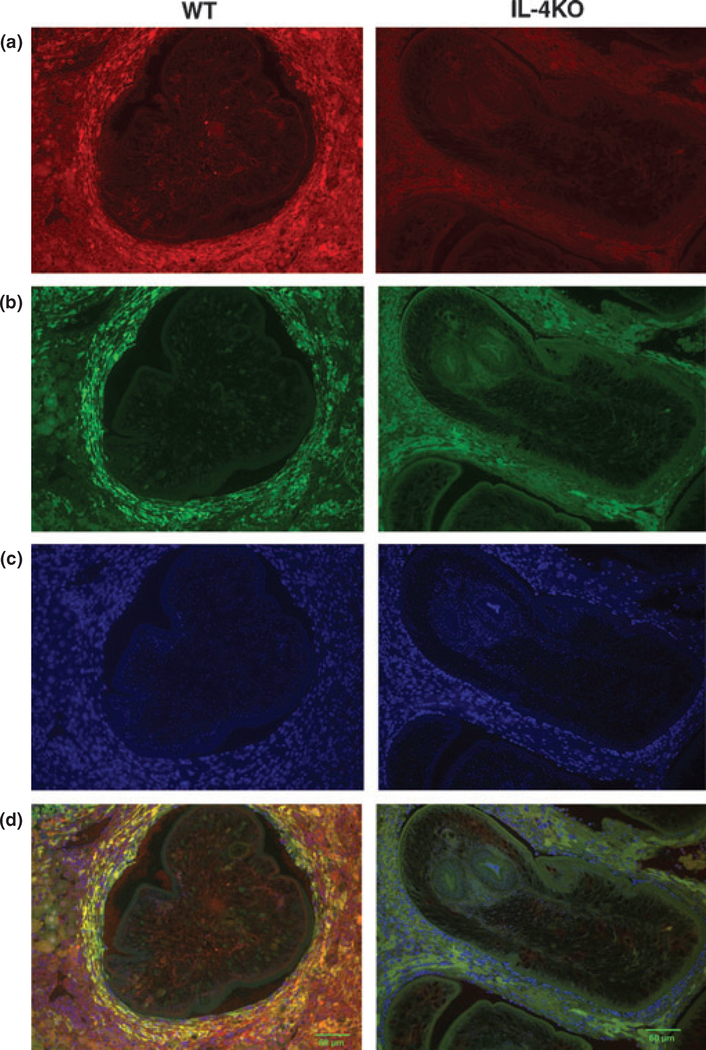
Protection against Mesocestoides corti, a cestode that invades vital organs, is dependent on the production of IL-4, as IL-4(-/-) mice were found to have higher parasite burdens when compared with wild-type mice. The goal of this study was to investigate the role of IL-4 in immunity to M. corti, focusing on the immunological profile and on potential mediators of pathology. IL-4(-/-) mice infected with M. corti showed 100% mortality by 32 days, whereas wild-type mice survived for approximately 1 year. Parasite burdens were significantly increased in the liver, peritoneal, and thoracic cavities of IL-4(-/-) mice, associated with impaired recruitment of inflammatory cells and a reduction in monocytes and macrophages. IL-5 production by splenocytes and expression in liver tissue was decreased in infected IL-4(-/-) mice compared with wild-type mice. In contrast, IL-4(-/-) mice produced increased amounts of IFNgamma and TNFalpha. Alternatively activated macrophages were a major feature of liver granulomas in wild-type mice evidenced by Arginase I expression, while livers from infected IL-4(-/-) mice showed impaired alternative macrophage activation without increased classical macrophage activation. Thus, lethality during M. corti infection of IL-4(-/-) mice is associated with decreased Th2 cytokines, increased Th1 cytokines and impairment of alternatively activated macrophages.


Figures

Figure 1
Figure 1
Lethal Mesocestoides corti infection in IL-4−/− mice is associated with decreased numbers of host inflammatory cells. C57BL/6 and IL-4−/− mice were infected intraperitoneally with approximately 600 M. corti tetrathyridia and followed for 32 days (a). The number of peritoneal exudate cells was determined (b). The total number of neutrophils, lymphocytes, monocytes/macrophages and eosinophils within the peritoneal exudate cell population was determined. (c) Data are a compilation of four experiments with equal numbers of wild-type and knock out mice. Asterisks indicate statistical difference from C57BL/6 mice as determined by anova (P < 0·05).
Figure 2
Figure 2
Stimulation of spleen cells from Mesocestoides corti infected C57BL/6 and IL-4−/− mice. Production of IL-5 (a) and IFNγ (b) following spleen cell stimulation with media, M. corti somatic antigen, or anti-CD3 antibody was measured by ELISA. The data displayed as mean ± SD and are representative of two experiments with each group containing 3–5 samples. A bar across a set indicates all groups below bar were statistically different from wild-type mice. Asterisks indicate statistical difference between IL-4−/− mice and C57BL/6 mice as determined by anova (P < 0·05).
Figure 3
Figure 3
Expression of liver cytokine mRNA from Mesocestoides corti infected C57BL/6 and IL-4−/− mice. Liver mRNA from M. corti infected C57BL/6 and IL-4 KO mice was isolated and reverse transcribed into cDNA, then analyzed by quantitative PCR. Shown are the relative quantities of IL-5, IL-13, TNFα, and IFNα gene expression after normalization to β-actin and standardization of the relative amount against a day 0 C57BL/6 sample. Statistical differences between IL-4−/− and C57BL/6 mice at a given time point are indicated by asterisks (P < 0·05 as determined by anova).
Figure 4
Figure 4
Histology of livers from C57BL/6 and IL-4−/− mice following infection with Mesocestoides corti. Liver samples from 14, 21, and 28 days post-infection in C57BL/6 wild-type and IL-4KO mice, displayed at low magnification (40×), demonstrate levels of parasite infiltration (a). Sections selected are representative of other sections prepared from the same mouse strain and time point. Levels of fibrosis in each liver sample were graded in a blinded fashion (b). The displayed results are the averages of five mice for each time point, and grading was done on liver regions which were representative of a median section of parasite infiltration. Data are displayed as mean ± SD.
Figure 5
Figure 5
Expression of mRNA for macrophage markers in the livers from C57BL/6 and IL-4−/− mice infected with Mesocestoides corti. Liver mRNA from M. corti infected C57BL/6 and IL-4−/− mice was isolated and reverse transcribed into cDNA, then analyzed by quantitative PCR. Shown are the relative quantities of Fizz-1 (Retnla), Ym-1 (Chi3l3), Arginase 1 (Arg 1) and iNOS (NOS2) gene expression after normalization to b actin and standardization of the relative amount against a day 0 C57BL/6 sample. Statistical differences between IL-4−/− and C57BL/6 mice at a given time point are indicated by asterisks (P < 0·05 as determined by anova).
Figure 6
Figure 6
IL-4-dependent alternatively activated macrophages surround Mesocestoides corti in liver. Immunohistochemical detection of F4/80 ± macrophages (a) (shown in red) surrounding M. corti larvae in liver at day 14 post-inoculation in C57BL/6 and IL-4−/− mice. Arginase-1 (b) was used as a marker for alternatively activated macrophages (shown in green) and cell nuclei (c) were stained with DAPI (shown in blue). Merged image (d) demonstrates Arginase-1 co-localization in macrophages in C57BL/6, but not in IL-4−/− mice. Representative photos of 50 granulomas examined per strain.

References

    1. Pollacco S, Nicholas WL, Mitchell GF & Stewart AC. T-cell dependent collagenous encapsulating response in the mouse liver to Mesocestoides corti (Cestoda). Int J Parasitol 1978; 8: 457–462. - PubMed
    1. Specht D & Widmer EA. Response of mouse liver to infection with tetrathyridia of Mesocestoides (Cestoda). J Parasitol 1972; 58: 431–437. - PubMed
    1. Rawat J, Dixon JB, Macintyre AR, McGarry HF & Taylor MJ. IL-4 dependent resistance to the tapeworm Mesocestoides corti (Cestoda) in mice. Parasite Immunol 2003; 25: 553–557. - PubMed
    1. Reed SG & Scott P. T-cell and cytokine responses in leishmaniasis. Curr Opin Immunol 1993; 5: 524–531. - PubMed
    1. Urban JF Jr, Madden KB, Svetic A, et al. The importance of Th2 cytokines in protective immunity to nematodes. Immunol Rev 1992; 127: 205–220. - PubMed

MeSH terms