. 2009;11(6):R78.
doi: 10.1186/bcr2445. Epub 2009 Nov 9.

A selective eradication of human nonhereditary breast cancer cells by phenanthridine-derived polyADP-ribose polymerase inhibitors

Affiliations

A selective eradication of human nonhereditary breast cancer cells by phenanthridine-derived polyADP-ribose polymerase inhibitors

Dana Inbar-Rozensal et al. Breast Cancer Res. 2009.

Abstract

Introduction: PARP-1 (polyADP-ribose polymerase-1) is known to be activated in response to DNA damage, and activated PARP-1 promotes DNA repair. However, a recently disclosed alternative mechanism of PARP-1 activation by phosphorylated externally regulated kinase (ERK) implicates PARP-1 in a vast number of signal-transduction networks in the cell. Here, PARP-1 activation was examined for its possible effects on cell proliferation in both normal and malignant cells.

Methods: In vitro (cell cultures) and in vivo (xenotransplants) experiments were performed.

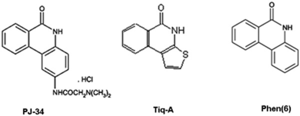
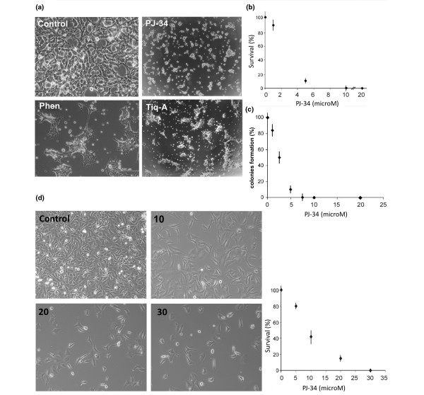
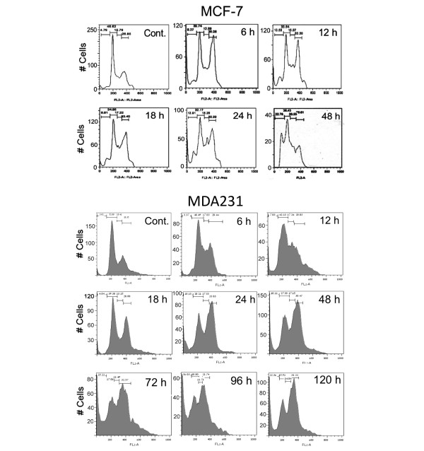
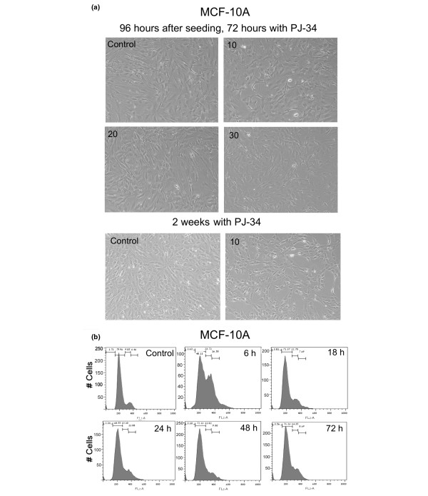
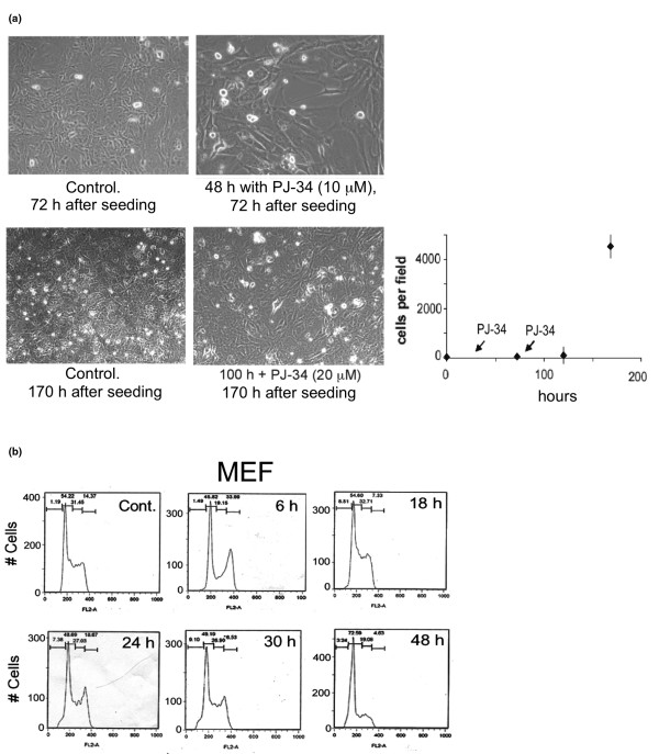
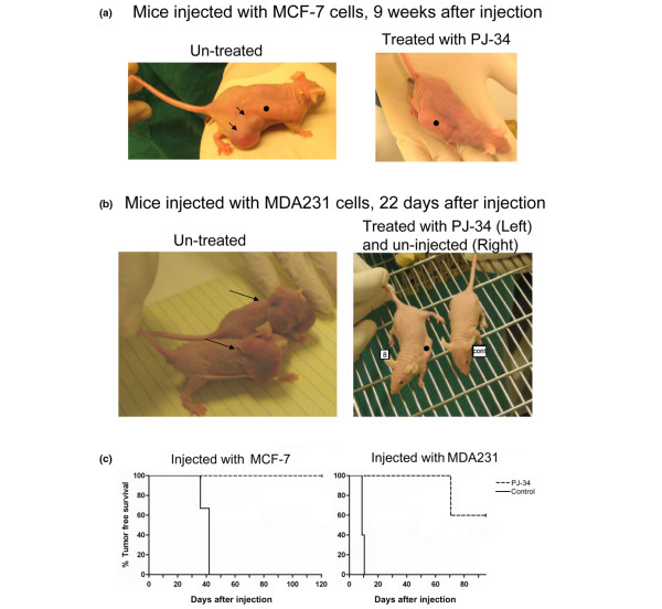
Results: Phenanthridine-derived PARP inhibitors interfered with cell proliferation by causing G2/M arrest in both normal (human epithelial cells MCF10A and mouse embryonic fibroblasts) and human breast cancer cells MCF-7 and MDA231. However, whereas the normal cells were only transiently arrested, G2/M arrest in the malignant breast cancer cells was permanent and was accompanied by a massive cell death. In accordance, treatment with a phenanthridine-derived PARP inhibitor prevented the development of MCF-7 and MDA231 xenotransplants in female nude mice. Quiescent cells (neurons and cardiomyocytes) are not impaired by these PARP inhibitors.

Conclusions: These results outline a new therapeutic approach for a selective eradication of abundant nonhereditary human breast cancers.


Figures

Figure 1
Figure 1
The chemical structures of the PARP inhibitors PJ-34, Tiq-A, and Phen.
Figure 2
Figure 2
Eradication of MCF-7 and MDA231 human breast cancer cells by phenanthridine-derived PARP inhibitors. (a) MCF-7 cells were seeded (about 500,000/well) in six-well plates and incubated for 48 hours with the potent PARP inhibitors, PJ-34 (10 μM), Tiq-A (50 μM), and Phen (25 μM), each applied (a single application) 24 hours after seeding. Cells were counted and pictured under a microscope (0.02 to 0.03 mm2 per field). Control (upper left) and treated MCF-7 cells are presented, 72 hours after seeding. These representative results were repeated in five different experiments. (b) Survival rate (percentage relative to untreated cells at each time point, control) of MCF-7 breast cancer cells after incubation for 48 hours with several concentrations of PJ-34, applied (a single application) 24 hours after seeding. Each value represents the average value of five measurements in different cell cultures. (c) Colony formation (percentage relative to colony formation of untreated cells) of MCF-7 breast cancer cells reseeded and incubated for 2 additional weeks in the absence of PJ-34, 48 hours after a single application of PJ-34 at several concentrations was applied 24 hours after the initial seeding (Methods). Each value represents the average value of three measurements in three different experiments. (d) Left panel: MDA231 breast cancer cells were seeded (about 500,000/well) in six-well plates. PJ-34 was applied into the medium at the indicated final concentrations 24 hours after seeding. Cells were counted and pictured under microscope (0.02 to 0.03 mm2 per field). Right panel: Survival rate (percentage relative to untreated cells at each time point, control) of MDA231 breast cancer cells after 72 hours of incubation with several concentrations of PJ-34, applied (a single application) 24 hours after seeding. Each value is an average of three measurements obtained in three different experiments.
Figure 3
Figure 3
G2/M arrest and cell death in MCF-7 and MDA231 treated with PJ-34. The effect of PJ-34 on the cell cycle was examined with flow cytometry. PJ-34 (10 μM) was applied to both types of cells, 24 hours after seeding. Controls: Untreated MCF-7 48 hours after seeding and untreated MDA231 cells 72 hours after seeding. At the indicated time, cells were collected, permeabilized (75% ethanol in DDW), and stained with propidium iodide (PI). The effects of PJ-34 on cell eradication and the kinetics of S-phase entry and G2/M transition were evaluated by the percentages of cells at these phases. G2/M arrest accompanied by cell death was detected in both cell types after 6 hours of incubation with PJ-34. Similar results for both cell types were obtained in three different experiments.
Figure 4
Figure 4
PJ-34 did not eradicate normal human epithelial cells MCF-10A. (a) MCF-10A cells were seeded (about 500,000/well) in six-well plates (Methods). PJ-34 was applied once at the indicated concentrations to MCF-10A cells 24 hours after seeding. Untreated MCF-10A cells and MCF-10 cells incubated with 10, 20, and 30 μM PJ-34 for 72 hours, and MCF-10A cells incubated with 10 μM PJ-34 for 2 weeks, were pictured under a microscope. These representative results were observed in three different experiments. (b) MCF-10A cells overcame G2/M arrest induced by treatment with PJ-34 (10 μM). Cells were analyzed with flow cytometry at the indicated periods after addition of 10 μM PJ-34. G2/M arrest detected 6 hours after PJ-34 application was relieved after 18 hours. Control represents untreated MCF-10A cells 72 hours after seeding. Similar results were measured in three different experiments.
Figure 5
Figure 5
PJ-34 did not eradicate mouse embryonic fibroblasts. (a) The effect of PJ-34 on mouse embryonic fibroblasts (MEF) was measured after repeated applications of PJ-34 (10 μM) 24 hours and 72 hours after seeding, as indicated. MEF, untreated and incubated with PJ-34 (10 μM) for 48 hours, and MEF, untreated and incubated with 20 μM PJ-34 for 100 hours are shown. On the right are displayed cell counts (0.02 to 0.03 mm2 per field) of cells incubated with PJ-34 (10 μM, after the first application, and 20 μM, after the second application) for the indicated time periods. Each value is an average value of cells counted in five different experiments. (b) MEFs were analyzed with flow cytometry at the indicated periods after addition of PJ-34. A transient G2/M transition arrest is indicated in MEFs incubated for 6 hours with PJ-34 (10 μM). Control: Untreated MEFs 48 hours after seeding. Similar results were obtained in three different experiments.
Figure 6
Figure 6
Treatment with PJ-34 prevented the development of MCF-7 and MDA231 xenotransplants. (a) Xenotransplants of MCF-7 developed within 6 to 7 weeks in three mice that were not treated with PJ-34 (control mice; tumors are indicated by arrows; left). Tumors did not develop in any of the three mice treated with PJ-34 for 14 days by a slow release of PJ-34 from a subcutaneously implanted osmotic pump (Alzet; pump •; Methods) (Right). Tumors were not detected during 4 months after injection with the MCF-7 cells. Each female CD-1 nu/nu mouse was injected with about 107 MCF-7 cells collected from 80% to 90% confluent cell cultures. Cells were immersed in Matrigel/PBS (Methods). Cells were injected near the pump, one hour after pump implantation. (b) Each CD-1 nu/nu female mouse was injected with about 107 MDA231 cells collected from 80% to 90% confluent cell cultures and immersed in Matrigel/PBS (Methods). Cells were injected near the pump, 24 hours after pump implantation. Xenotransplants of MDA231 developed within 10 days in five female mice that were not treated with PJ-34 (untreated mice). Tumors did not develop in five female mice treated for 14 days with a slow release from a subcutaneously implanted osmotic pump (Alzet pump). Tumors were not detected in these mice during 10 weeks after injection with MDA231 cells and 8 weeks after the treatment with PJ-34. A detailed presentation of this experiment is included in Additional data file 1. (c) Kaplan-Meier survival analysis is used for presenting tumor-free survival curves of mice injected with MCF-7 cells and mice injected with MDA231 cells, without or after treatment with PJ-34, as described earlier. The significance (log-rank significance test) was P = 0.0253 for mice injected with MCF-7 cells, and P = 0.0023 for mice injected with MDA231 cells.

Comment in

References

    1. Schreiber V, Dantzer FJ-C, Ame de Murcia G. PolyADP-ribose: novel functions for an old molecule. Nat Rev Mol Cell Biol. 2006;7:517–528. doi: 10.1038/nrm1963. - DOI - PubMed
    1. Kraus WL. Transcriptional control by PARP-1:chromatin modulation, enhancer-binding, coregulation, and insulation. Curr Opin Cell Biol. 2008;20:294–302. doi: 10.1016/j.ceb.2008.03.006. - DOI - PMC - PubMed
    1. Cohen-Armon M, Visochek L, Rozensal D, Kalal A, Geistrikh I, Klein R, Bendetz-Nezer S, Yao Z, Seger R. DNA-independent PARP-1 activation by phosphorylated ERK2increases Elk1 activity: a link to histone acetylation. Mol Cell. 2007;25:297–308. doi: 10.1016/j.molcel.2006.12.012. - DOI - PubMed
    1. Cohen-Armon M. PARP-1 activation in the ERK signaling pathway. Trends Pharmacol Sci. 2007;28:556–560. doi: 10.1016/j.tips.2007.08.005. - DOI - PubMed
    1. Cohen-Armon M. PARP-1 activation mediates the expression of immediate early genes implicated in long-term memory formation. FEBS J. 2008;275(Suppl 1):92.

Publication types

MeSH terms