A hierarchical method based on active shape models and directed Hough transform for segmentation of noisy biomedical images; application in segmentation of pelvic X-ray images
- PMID: 19891796
- PMCID: PMC2773917
- DOI: 10.1186/1472-6947-9-S1-S2
A hierarchical method based on active shape models and directed Hough transform for segmentation of noisy biomedical images; application in segmentation of pelvic X-ray images
Abstract
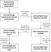
Background: Traumatic pelvic injuries are often associated with severe, life-threatening hemorrhage, and immediate medical treatment is therefore vital. However, patient prognosis depends heavily on the type, location and severity of the bone fracture, and the complexity of the pelvic structure presents diagnostic challenges. Automated fracture detection from initial patient X-ray images can assist physicians in rapid diagnosis and treatment, and a first and crucial step of such a method is to segment key bone structures within the pelvis; these structures can then be analyzed for specific fracture characteristics. Active Shape Model has been applied for this task in other bone structures but requires manual initialization by the user. This paper describes a algorithm for automatic initialization and segmentation of key pelvic structures - the iliac crests, pelvic ring, left and right pubis and femurs - using a hierarchical approach that combines directed Hough transform and Active Shape Models.
Results: Performance of the automated algorithm is compared with results obtained via manual initialization. An error measures is calculated based on the shapes detected with each method and the gold standard shapes. ANOVA results on these error measures show that the automated algorithm performs at least as well as the manual method. Visual inspection by two radiologists and one trauma surgeon also indicates generally accurate performance.
Conclusion: The hierarchical algorithm described in this paper automatically detects and segments key structures from pelvic X-rays. Unlike various other x-ray segmentation methods, it does not require manual initialization or input. Moreover, it handles the inconsistencies between x-ray images in a clinical environment and performs successfully in the presence of fracture. This method and the segmentation results provide a valuable base for future work in fracture detection.
Figures


Similar articles
-
Unified wavelet and Gaussian filtering for segmentation of CT images; application in segmentation of bone in pelvic CT images.BMC Med Inform Decis Mak. 2009 Nov 3;9 Suppl 1(Suppl 1):S8. doi: 10.1186/1472-6947-9-S1-S8. BMC Med Inform Decis Mak. 2009. PMID: 19891802 Free PMC article.
-
A new hierarchical method for multi-level segmentation of bone in pelvic CT scans.Annu Int Conf IEEE Eng Med Biol Soc. 2011;2011:3399-402. doi: 10.1109/IEMBS.2011.6090920. Annu Int Conf IEEE Eng Med Biol Soc. 2011. PMID: 22255069
-
Fully automatic detection of salient features in 3-d transesophageal images.Ultrasound Med Biol. 2014 Dec;40(12):2868-84. doi: 10.1016/j.ultrasmedbio.2014.07.014. Epub 2014 Oct 11. Ultrasound Med Biol. 2014. PMID: 25308940
-
A semiautomatic segmentation method framework for pelvic bone tumors based on CT-MR multimodal images.Int J Numer Method Biomed Eng. 2023 Oct;39(10):e3697. doi: 10.1002/cnm.3697. Epub 2023 Mar 31. Int J Numer Method Biomed Eng. 2023. PMID: 36999653
-
Vertebra identification using template matching modelmp and K-means clustering.Int J Comput Assist Radiol Surg. 2014 Mar;9(2):177-87. doi: 10.1007/s11548-013-0927-2. Epub 2013 Jul 24. Int J Comput Assist Radiol Surg. 2014. PMID: 23881250 Review.
Cited by
-
Automated assessment of pelvic radiographs using deep learning: A reliable diagnostic tool for pelvic malalignment.Heliyon. 2024 Apr 16;10(8):e29677. doi: 10.1016/j.heliyon.2024.e29677. eCollection 2024 Apr 30. Heliyon. 2024. PMID: 38660256 Free PMC article.
-
An Automated Deep Learning Method for Tile AO/OTA Pelvic Fracture Severity Grading from Trauma whole-Body CT.J Digit Imaging. 2021 Feb;34(1):53-65. doi: 10.1007/s10278-020-00399-x. Epub 2021 Jan 21. J Digit Imaging. 2021. PMID: 33479859 Free PMC article.
-
Quantification and Characterization of CTCs and Clusters in Pancreatic Cancer by Means of the Hough Transform Algorithm.Int J Mol Sci. 2023 Feb 21;24(5):4278. doi: 10.3390/ijms24054278. Int J Mol Sci. 2023. PMID: 36901704 Free PMC article.
References
-
- Basta A, Blackmore C, Wessells H. Predicting urethral injury from pelvic fracture patterns in male patients with blunt trauma. J Urology. 2007;177:571–575. - PubMed
-
- Schmal H, Markmiller M, Mehlhorn A, Sudkamp N. Epidemiology and outcome of complex pelvic injury. Acta Orthopaedica Belgica. 2005;71 - PubMed
-
- Tan S, Porter K. Free fall trauma. J Trauma. 2006;8:157–167.
-
- ACOS . ATLS Advanced Trauma Life Support Program for Doctors. 7. American College of Surgeons; 2004.
-
- Haas B, Coradi T, Scholz M, Kunz P, Huber M, Oppitz U, Andre L, Lengkeek V, Huyskens D, van Esch A, Reddick R. Automatic segmentation of thoracic and pelvic CT images for radiotherapy planning using implicit anatomic knowledge and organ-specific segmentation strategies. Phys Med Biol. 2008;53:1751–71. - PubMed
Publication types
MeSH terms
LinkOut - more resources
Full Text Sources

