. 2009 Nov 10;54(20):1891-8.
doi: 10.1016/j.jacc.2009.07.031.

Substrate-specific derangements in mitochondrial metabolism and redox balance in the atrium of the type 2 diabetic human heart

Affiliations

Substrate-specific derangements in mitochondrial metabolism and redox balance in the atrium of the type 2 diabetic human heart

Ethan J Anderson et al. J Am Coll Cardiol. .

Abstract

Objectives: The aim of this study was to determine the impact of diabetes on oxidant balance and mitochondrial metabolism of carbohydrate- and lipid-based substrates in myocardium of type 2 diabetic patients.

Background: Heart failure represents a major cause of death among diabetic patients. It has been proposed that derangements in cardiac metabolism and oxidative stress may underlie the progression of this comorbidity, but scarce evidence exists in support of this mechanism in humans.

Methods: Mitochondrial oxygen (O(2)) consumption and hydrogen peroxide (H(2)O(2)) emission were measured in permeabilized myofibers prepared from samples of the right atrial appendage obtained from nondiabetic (n = 13) and diabetic (n = 11) patients undergoing nonemergent coronary artery bypass graft surgery.

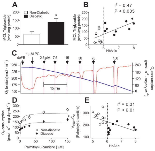
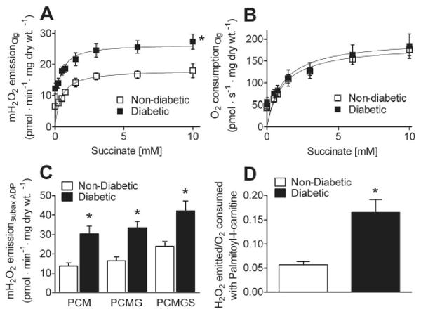
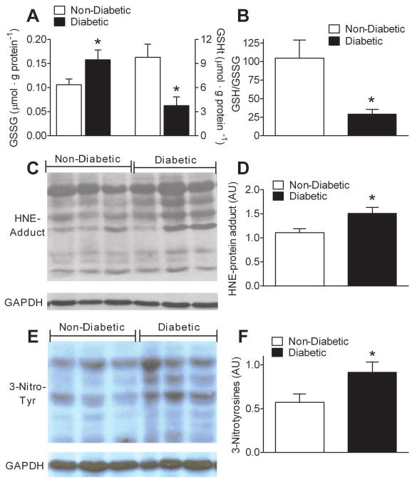
Results: Mitochondria in atrial tissue of type 2 diabetic individuals show a sharply decreased capacity for glutamate and fatty acid-supported respiration, in addition to an increased content of myocardial triglycerides, as compared to nondiabetic patients. Furthermore, diabetic patients show an increased mitochondrial H(2)O(2) emission during oxidation of carbohydrate- and lipid-based substrates, depleted glutathione, and evidence of persistent oxidative stress in their atrial tissue.

Conclusions: These findings are the first to directly investigate the effects of type 2 diabetes on a panoply of mitochondrial functions in the human myocardium using cellular and molecular approaches, and they show that mitochondria in diabetic human hearts have specific impairments in maximal capacity to oxidize fatty acids and glutamate, yet increased mitochondrial H(2)O(2) emission, providing insight into the role of mitochondrial dysfunction and oxidative stress in the pathogenesis of heart failure in diabetic patients.


Conflict of interest statement

Disclosures: The authors have no conflicts of interest with industry to disclose.

Figures

Figure 1
Figure 1. High levels of IMCL triglycerides in atrium of type 2 diabetic patients is linked to reduced maximal capacity of mitochondrial fatty acid oxidation
A, Levels of IMCL triglycerides in atrial tissue homogenate prepared from type 2 Diabetic and Non-Diabetic patients. B, Plot showing correlation between myocardial triglycerides and HbA1c in Diabetic (○) and Non-Diabetic (●) patients (B). Shown in C is a representative trace of ADP-stimulated O2 consumption in permeabilized human atrial myofibers in response to incrementally increasing concentrations of palmitoyl-L-carnitine. Permeabilized fibers in the absence of substrate (deFB) were added to respiratory medium in the presence of 3 mM ADP, 5 mM glucose, 1 U/ml hexokinase (to create permanent, maximally phosphorylating respiratory state), followed by incrementally increasing concentrations of palmitoyl-L-carnitine (PC) in the presence of 2 mM malate. The blue line is O2 concenctration in the medium (left Y-axis), the red line is rate of O2 consumption (right Y-axis). D, Kinetic plots of palmitoyl-L-carnitine supported respiration (i.e. fatty acid oxidation) in permeabilized atrial myofibers prepared from both groups of patients, and the correlation of Vmax with HbA1c (E). Quantified data are means ± S.E.M, N = 9-12 patients in each group. * P < 0.05 vs Non-Diabetic.
Figure 2
Figure 2. Glutamate-specific impairment in maximal respiratory capacity in atrium of type 2 diabetic patients
A, Kinetic plots of ADP-stimulated respiration supported by 10 mM pyruvate and 2 mM malate in permeabilized atrial myofibers prepared from Diabetic and Non-Diabetic patients. B, Minimal (S0) and maximal (SADP) respiration supported by succinate in both groups. C, representative trace of a typical O2 consumption experiment in permeabilized human atrial myofibers using sequential addition of oxidative substrates, nucleotides and inhibitors to assess contribution of multiple oxidative substrates to total respiratory flux. Permeabilized fibers in the absence of substrate were added to respiratory medium (deFB), followed by 5 mM glutamate/2 mM malate (GM0), 5 mM ADP (GMADP), 10 mM succinate (GMSADP), 20 μM cytochrome C (to test for intactness of outer mitochondrial membrane), 10 μg/ml oligomycin (GMSO), and 3 μM FCCP (GMSF). D, Quantified rates of glutamate-supported respiration in permeabilized atrial myofibers of Diabetic and Non-Diabetic patients. Quantified data are means ± S.E.M, N = 7-8 patients in each group. * P < 0.05 vs Non-Diabetic.
Figure 3
Figure 3. Mitochondria in atrium of type 2 diabetic patients display high levels of mitochondrial H2O2 emission while oxidizing carbohydrate- and lipid-based substrates
A, Kinetic plots of mitochondrial H2O2 emission and (B) O2 consumption in permeabilized atrial myofibers prepared from Diabetic and Non-Diabetic patients supported by incrementally increasing concentrations of succinate in the presence of 10 μg/ml of oligomycin (to inhibit ATPase and ensure basal respiratory state) + 5 mM glutamate, 2 mM malate. C, Quantified rates of mitochondrial H2O2 emission during respiration in the presence of 125 μM ADP, 5 mM glucose, 1 U/ml hexokinase (to create permanent, submaximally phosphorylating respiratory state), supported by 75μM palmitol-L-carnitine + 2 mM malate (PCM), 5 mM glutamate (PCMG) and 10 mM succinate (PCMGS). D, Ratio of moles of H2O2 emitted per mole of O2 consumed during respiration supported by palmitoyl-L-carnitine. Quantified data are means ± S.E.M, N = 7-9 patients in each group. * P < 0.05 vs Non-Diabetic.
Figure 4
Figure 4. Altered glutathione redox status and evidence of persistent lipo-peroxyl and nitrosative stress in atrial tissue of type 2 diabetic patients
A, Quantified levels of oxidized glutathione (GSSG), and total glutathione (GSHt), in atrial tissue of Diabetic and Non-Diabetic patients. B, Redox environment of atrial tissue as determined by GSH/GSSG ratio. C, Representative immunoblot and D, densitometric quantification of hydroxynonenal (HNE)-modified proteins in atrial tissue from both groups of patients. E, Representative immunoblot and F, densitometric quantification of 3-nitrotyrosine-modified proteins in atrial tissue from both groups of patients. Quantified data are means ± S.E.M, N = 8-11 patients in each group. * P < 0.05 vs Non-Diabetic.

Comment in

References

    1. Boudina S, Abel ED. Diabetic cardiomyopathy revisited. Circulation. 2007;115:3213–23. - PubMed
    1. Zhou YT, Grayburn P, Karim A, et al. Lipotoxic heart disease in obese rats: implications for human obesity. Proc Natl Acad Sci U S A. 2000;97:1784–9. - PMC - PubMed
    1. Saddik M, Lopaschuk GD. Triacylglycerol turnover in isolated working hearts of acutely diabetic rats. Can J Physiol Pharmacol. 1994;72:1110–9. - PubMed
    1. McGavock JM, Lingvay I, Zib I, et al. Cardiac steatosis in diabetes mellitus: a 1H-magnetic resonance spectroscopy study. Circulation. 2007;116:1170–5. - PubMed
    1. Rijzewijk LJ, van der Meer RW, Smit JW, et al. Myocardial steatosis is an independent predictor of diastolic dysfunction in type 2 diabetes mellitus. J Am Coll Cardiol. 2008;52:1793–9. - PubMed

Publication types

MeSH terms