. 2010 Jan;38(2):441-54.
doi: 10.1093/nar/gkp905. Epub 2009 Nov 5.

The Mre11/Rad50/Nbs1 complex functions in resection-based DNA end joining in Xenopus laevis

Affiliations

The Mre11/Rad50/Nbs1 complex functions in resection-based DNA end joining in Xenopus laevis

Elaine M Taylor et al. Nucleic Acids Res. 2010 Jan.

Abstract

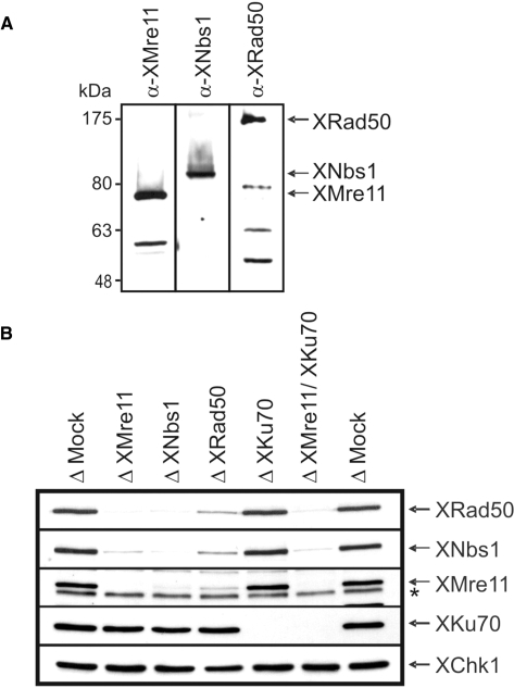
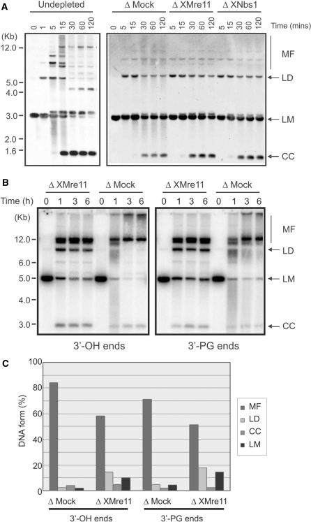
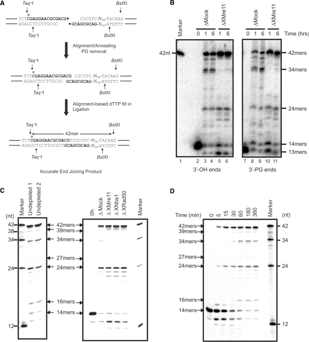
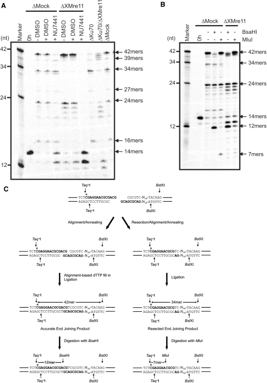
The repair of DNA double-strand breaks (DSBs) is essential to maintain genomic integrity. In higher eukaryotes, DNA DSBs are predominantly repaired by non-homologous end joining (NHEJ), but DNA ends can also be joined by an alternative error-prone mechanism termed microhomology-mediated end joining (MMEJ). In MMEJ, the repair of DNA breaks is mediated by annealing at regions of microhomology and is always associated with deletions at the break site. In budding yeast, the Mre11/Rad5/Xrs2 complex has been demonstrated to play a role in both classical NHEJ and MMEJ, but the involvement of the analogous MRE11/RAD50/NBS1 (MRN) complex in end joining in higher eukaryotes is less certain. Here we demonstrate that in Xenopus laevis egg extracts, the MRN complex is not required for classical DNA-PK-dependent NHEJ. However, the XMRN complex is necessary for resection-based end joining of mismatched DNA ends. This XMRN-dependent end joining process is independent of the core NHEJ components Ku70 and DNA-PK, occurs with delayed kinetics relative to classical NHEJ and brings about repair at sites of microhomology. These data indicate a role for the X. laevis MRN complex in MMEJ.


Figures

Figure 1.
Figure 1.
Immunodepletion of the MRN complex and Ku70 from X. laevis egg extract. (A) Xenopus laevis egg extract was analysed by Western blotting with antisera against XMre11, XNbs1 and XRad50. (B) Extract was subjected to three rounds of depletion using protein A sepharose coupled to non-specific rabbit IgGs (Δ Mock), anti-XMre11 antibodies (Δ XMre11), anti-XNbs1 antibodies (Δ XNbs1) or anti-XRad50 antibodies (Δ XRad50) or using protein G sepharose coupled to antibodies against Ku70 (Δ XKu70). Double depletion of XMre11 and XKu70 was achieved by two rounds of depletion with α-Ku70 beads followed by one round with α-XMre11 beads. Depletion efficiency was analysed by Western blotting with the appropriate antisera. XChk1 was used as a loading control. A non-specific band is denoted by asterisk.
Figure 2.
Figure 2.
NHEJ in XMRN-depleted extracts. (A) Southern blot analysis of NHEJ products generated by incubation of linearized pUC19 template (1 ng/μl) in the indicated egg extracts. Repair products were detected using a fluorescein-labelled pUC19 probe. LMs were converted into MFs, linear dimers (LDs) and closed circular DNA monomers (CC). (B) Agarose gel analysis of repair products resulting from the incubation of radio-labelled pSV56 linear substrates with defined termini in mock-depleted (Δ Mock) or XMre11-depleted (Δ XMre11) extract. Left panel shows end joining of 3′-OH ends. Right panel shows repair of 3′-PG termini. (C) Quantification of DNA forms after 6 h end joining reactions using pSV56 defined linear substrates. The amount of each DNA form is expressed as a percentage of the total signal for each individual lane.
Figure 3.
Figure 3.
Analysis of DNA end joining at the nucleotide level. (A) Formation of accurate end joining products from 3′-OH or 3′-PG end substrates during NHEJ reactions. Following the removal of the 3′-PG moeity (•), the 3′-terminal CG residues align and the one-base gap opposite A is filled in by a DNA polymerase. Ligation of the ends generates accurate end joined products which, following treatment with the restriction enzymes TaqαI and BstXI, can be observed on a sequencing gel as 42-bp oligomers. (B) Sequencing gel analysis of NHEJ in X. laevis egg extracts using 3′-OH- and 3′-PG-defined substrates. pSV56 linear substrates with either 3′-OH or 3′-PG ends (1 ng/μl) were incubated in mock-depleted (Δ Mock) or XMre11-depleted (Δ XMre11) extract at 21°C for the indicated times. DNA was digested with TaqαI and BstXI, then resolved on a 20% denaturing polyacrylamide gel. (C) Linearized pSV56 with 3′-OH termini was incubated at 1 ng/μl in undepleted, mock-depleted and XMre11-, XNbs1- or XRad50-depleted extracts for 6 h at 21°C before digestion and analysis as described above. Substrate added to mock-depleted extract at 1 ng/μl and processed immediately serves as a control (0 h). (D) Linearized pSV56 with 3′-OH ends was incubated at 1 ng/μl in X. laevis egg extract at 21°C for the indicated times. Accurate repair occurs at earlier time points than resection-based repair.
Figure 4.
Figure 4.
Resection-based end joining is Ku70 independent and occurs at sites of microhomology. (A) pSV56 3′-OH substrate was incubated in mock-, XMre11-, Ku70- and XMre11/Ku70 double-depleted extracts for 6 h. DMSO was added to a final concentration of 0.4% and NU7441 was added to a final concentration of 8 µM as indicated. (B) pSV56 3′-OH substrate was incubated in mock- or XMre11-depleted extract at 21°C for 6 h. DNA was cut with TaqαI and BstXI then further digested with BsaHI or MluI as indicated. (C) Alignment of the 3′-terminal CG residues, dTTP fill-in and ligation generates a BsaHI restriction site (5′-GRCGYC-3′) 12-bp downstream of the TaqαI site in the accurately repaired 42-mer. The 3′–5′ resection of 7 bp on either side of the DNA break creates 4-bp CGCG overhangs that anneal and are ligated without a requirement for further synthesis to yield the 34-mer resected end joining product. A diagnostic MluI restriction site (ACGCGT) is generated 7-bp downstream of the TaqαI site by repair in this manner.

References

    1. Khanna KK, Jackson SP. DNA double-strand breaks: signaling, repair and the cancer connection. Nat. Genet. 2001;27:247–254. - PubMed
    1. van Gent DC, Hoeijmakers JH, Kanaar R. Chromosomal stability and the DNA double-stranded break connection. Nat. Rev. 2001;2:196–206. - PubMed
    1. Symington LS. Role of RAD52 epistasis group genes in homologous recombination and double-strand break repair. Microbiol. Mol. Biol. Rev. 2002;66:630–670. - PMC - PubMed
    1. Jazayeri A, Falck J, Lukas C, Bartek J, Smith GC, Lukas J, Jackson SP. ATM- and cell cycle-dependent regulation of ATR in response to DNA double-strand breaks. Nat. Cell. Biol. 2006;8:37–45. - PubMed
    1. West SC. Molecular views of recombination proteins and their control. Nat. Rev. Mol. Cell. Biol. 2003;4:435–445. - PubMed

Publication types

MeSH terms