. 2009 Nov 6;326(5954):853-8.
doi: 10.1126/science.1177585.

Small-molecule activators of a proenzyme

Affiliations

Small-molecule activators of a proenzyme

Dennis W Wolan et al. Science. .

Abstract

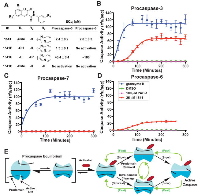
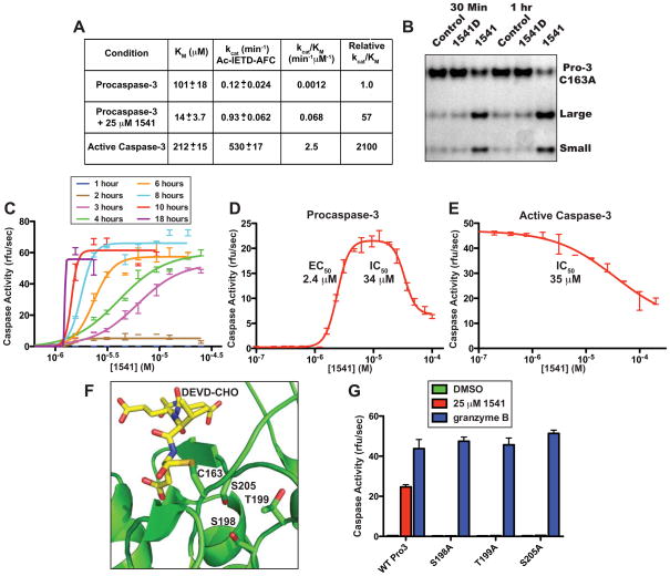
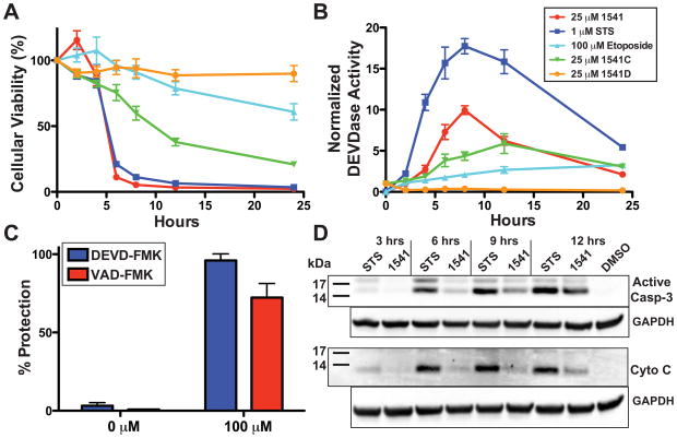
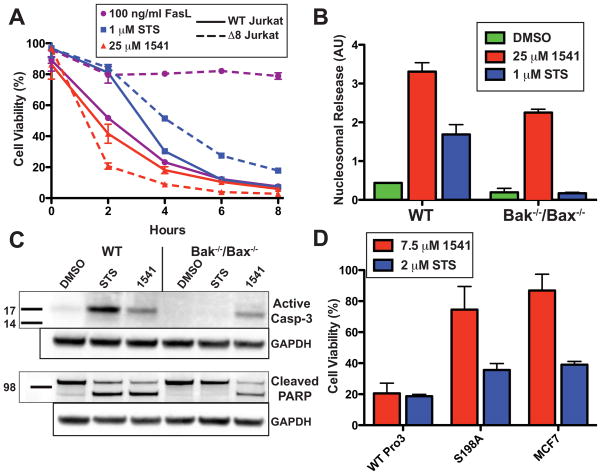
Virtually all of the 560 human proteases are stored as inactive proenyzmes and are strictly regulated. We report the identification and characterization of the first small molecules that directly activate proenzymes, the apoptotic procaspases-3 and -6. It is surprising that these compounds induce autoproteolytic activation by stabilizing a conformation that is both more active and more susceptible to intermolecular proteolysis. These procaspase activators bypass the normal upstream proapoptotic signaling cascades and induce rapid apoptosis in a variety of cell lines. Systematic biochemical and biophysical analyses identified a cluster of mutations in procaspase-3 that resist small-molecule activation both in vitro and in cells. Compounds that induce gain of function are rare, and the activators reported here will enable direct control of the executioner caspases in apoptosis and in cellular differentiation. More generally, these studies presage the discovery of other proenzyme activators to explore fundamental processes of proenzyme activation and their fate-determining roles in biology.


Figures

Fig. 1
Fig. 1
Specificity of small molecule activators for executioner procaspases. (A) Chemical analogs of 1541 and EC50 values of procaspases-3 and -6 activation. Small molecule activators require the imidazopyridine moiety and prefer a heteroatom substituent at the 8-position on the coumarin ring (39)(40). Substitution of the 8-methoxy with an 8-hydroxy (1541B) improves the EC50 and specificity for activation of procaspase-3 over procaspase-6. Addition of a 6-bromo (1541C) reduces potency to both procaspases. Complete removal of the imidazopyridine substituent (1541D) ablates activation of procaspases-3 and -6. (BD) Time course of executioner procaspase activation by 1541 facilitates full self-cleavage of procaspases-3 and -6. Procaspases-3 (B), -7 (C) and -6 (D) were incubated at 100 nM with 1 nM granzyme B (blue), DMSO (green on baseline) 100 μM PAC-1 (magenta) or 25 μM 1541 (red) at 37 °C and assayed for activity by addition of fluorogenic peptide substrates Ac-DEVD-AFC (caspases-3 and -7) or Ac-VEID-AFC (caspase-6) every 20 minutes (41). (E) Proposed model for small molecule-assisted procaspase self-activation. We hypothesize that procaspases are in a dynamic equilibrium between an off-state (left) and on-state (right), similar to mature caspases. Unlike mature caspases, the population favors the off-state conformation. Upon binding a small molecule activator, the equilibrium shifts to an on-state (center). This complex slowly undergoes autoproteolytic activation (black arrows) that accelerates with increasing production of mature caspase (green arrows). This model also accounts for activation at low concentrations of small molecule where the on-state is preferred and one active site is available in the dimer for processing, and, at high concentrations, where both sites are saturated and can lead to inhibition.
Fig. 2
Fig. 2
In vitro characterization of apoptotic procaspase small molecule activators. (A) Initial rates of activation of procaspase-3 in presence of 1541B, as measured by Ac-IETD-AFC compared to unstimulated zymogen and mature caspase-3. Procaspase-3 (100 nM) was incubated with 1541B (0.78 to 50 μM) and initial rates of activity were measured over ~15 min by addition of Ac-IETD-AFC (1.2 to 300 μM) at 37 °C. Presence of 1541 improves the kcat/KM of procaspase-3 by 57-fold and stimulates autocatalytic processing. (B) Silver-stained SDS-PAGE gel of inactive C163A procaspase-3 (250 nM) incubated with granzyme B (0.5 nM) with and without 1541 (25 μM) shows increased cleavage susceptibility by granzyme B with 1541 but not with the inactive analog 1541D. (C) Procaspase-3 was incubated at 100 nM with 1541 (0.1 to 100 μM) to assess rate of self-activation. Kinetic activity of the mixtures was determined every hour for 18 hours by incubation with 20 μM Ac-DEVD-AFC. Once procaspase-3 is activated by 1541, the zymogen rapidly self-activates. The effective concentration of 1541 is approximately 1.5 μM with no activation below this concentration despite extended incubation periods. (D) Procaspase-3 at 100 nM was incubated with 1541 (100 nM to 100 μM). After a 4-hour incubation at 37 °C, the samples were assayed for caspase activity by addition of Ac-DEVD-AFC. Low concentrations of 1541 induce activity of procaspase-3 with an EC50 of 2.4 μM, and, at high concentrations, inhibit with an IC50 of 34.0 μM. (E) 1541 exerts a similar inhibitory effect on active caspase-3 (IC50 = 35 μM), as seen for procaspase-3. Mature caspase-3 was incubated at 25 nM in various concentrations of 1541 for 10 minutes and assayed with Ac-DEVD-AFC. (F) Structural model of the active site of procaspase-3 showing the residues targeted for mutation. The location of the DEVD substrate is shown and modeled from caspase-3 in complex with Ac-DEVD-CHO (PDB 2DKO). (G) Identification of mutations in procaspase-3 that confer resistance to activation by 1541. Wild type and mutant (S198A, T199A and S205A) procaspases were incubated for 5 hours with 25 μM 1541 (red), 1 nM granzyme B (blue) or DMSO (green on baseline) at 37 °C and sampled for activation via kinetic assays initiated by addition of substrate Ac-DEVD-AFC. The procaspase-3 variants S198A, T199A and S205A displayed wild type activity when activated by granzyme B, but were unable to self-activate in the presence of 1541.
Fig. 3
Fig. 3
Induction of apoptosis in a p53-deficient breast cancer cell line by 1541. (A) Time course of cell viability in BT549 cells. 2,000 BT549 cells were incubated with either 25 μM 1541 (red), 25 μM 1541C (green), 25 μM 1541D (orange), 1 μM STS (blue), 100 μM etoposide (light blue), or DMSO (final 0.5%) and assayed for cell viability at 2, 4, 6, 8, 12 and 24 hours. Analogs 1541C and D induce limited to no cellular death, respectively, with correlation to the in vitro effects on procaspase-3 self-activation. (B) Time course of DEVDase activity in BT549 cells. All concentrations and variables are conserved with panel 3A. 1541 induces less DEVDase activity in comparison to STS, but results in similar rates of apoptosis. (C) Protection against 1541-induced apoptosis with caspase inhibitors. 2,500 cells from BT549 were incubated overnight with the caspase-3/-7 inhibitor Biotin-DEVD-FMK (blue) or the general caspase inhibitor Biotin-VAD-FMK (red) from 0.78 to 100 μM. Cells were exchanged into new media containing 25 μM 1541 and respective concentration of peptide inhibitor. After 24 hours of incubation at 37 °C with 1541, the cells were assayed for cell viability. Caspase inhibitors prevented cellular apoptosis induced by 1541. (D) Cytoplasmic extracts of BT549 cells reveal limited cytochrome C release from mitochondria during 1541-induced apoptosis. One million BT549 cells were incubated with 25 μM 1541, 1 μM STS, or DMSO (0.5% final) and harvested every 3 hours. Cytoplasmic supernatants were subjected to Western blot analysis for presence of active caspase-3 or cytochrome C. Compared to STS, 1541 induced far less release of cytochrome C during apoptosis.
Fig. 4
Fig. 4
Validation of executioner procaspases as cellular target of 1541. (A) Wild type (A3) (solid line) or caspase-8 deficient (I9.2) (dotted line) Jurkat cells (100,000 cells) were incubated with 25 μM 1541 (red), 1 μM STS (blue) or 100 ng/ml FasL (purple) and assayed for cellular viability every 2 hours. 1541 induces cell death independent of the extrinsic pathway. (B) 5,000 cells derived from either wild type or Bak−/−/Bax−/− DKO MEFs were incubated with either 1 μM STS (blue), 25 μM 1541 (red) or DMSO (green) (final 0.5%) at 37 °C. A histone sandwich ELISA (Roche) was performed on the lysates to determine the extent of apoptosis after 12 hours. Induction of cell death by 1541, in the absence of Bak and Bax, supports direct activation of the executioner procaspases. (C) For Western blot analysis, one million cells of both wild type and Bak−/−/Bax−/− DKO MEFs were plated and incubated with either 1 μM STS, 25 μM 1541 or DMSO (final 0.5%) at 37 °C for 12 hours. Western blots of whole-cell lysates were probed for the presence of active caspase-3 and cleavage of PARP. (D) Transfection of MCF-7 cells with wild type procaspase-3 and 1541-resistant S198A procaspase-3 supports ablation of 1541-induced procaspase-3 activation in cells. 6,000 cells from stably transfected MCF-7 cell lines were incubated for 24 hours with either 7.5 μM 1541 (red), 2 μM STS (blue) or DMSO (final concentration 0.5%) and assayed for cell viability. S198A procaspase-3 MCF-7 cells resisted apoptosis similarly to the parental MCF-7 cell line, while wild type procaspase-3 transfection of MCF-7 cells had increased apoptotic susceptibility to 1541.

References

    1. Neurath H, Walsh KA. Proc Natl Acad Sci USA. 1976;73:3825. - PMC - PubMed
    1. Lupardus PJ, Shen A, Bogyo M, Garcia KC. Science. 2008;322:265. - PMC - PubMed
    1. Stack MS, Pizzo SV. J Biol Chem. 1993;268:18924. - PubMed
    1. Earnshaw WC, Martins LM, Kaufmann SH. Annu Rev Biochem. 1999;68:383. - PubMed
    1. Yi CH, Yuan J. Dev Cell. 2009;16:21. - PMC - PubMed

Publication types

MeSH terms