Review
. 2009 Nov 6;105(10):934-47.
doi: 10.1161/CIRCRESAHA.109.201400.

Origin of cardiac fibroblasts and the role of periostin

Affiliations
Review

Origin of cardiac fibroblasts and the role of periostin

Paige Snider et al. Circ Res. .

Abstract

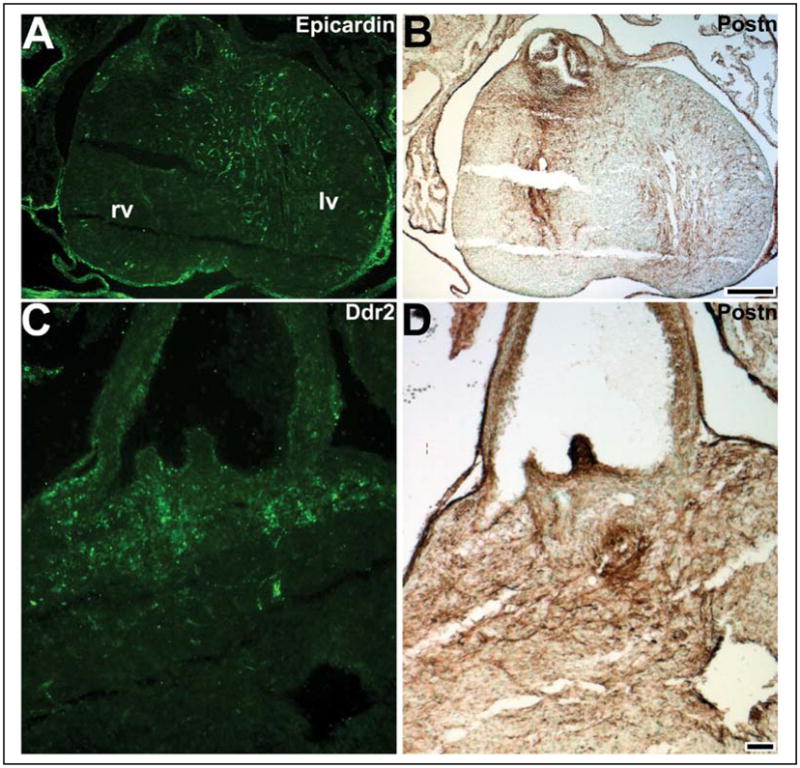
Cardiac fibroblasts are the most populous nonmyocyte cell type within the mature heart and are required for extracellular matrix synthesis and deposition, generation of the cardiac skeleton, and to electrically insulate the atria from the ventricles. Significantly, cardiac fibroblasts have also been shown to play an important role in cardiomyocyte growth and expansion of the ventricular chambers during heart development. Although there are currently no cardiac fibroblast-restricted molecular markers, it is generally envisaged that the majority of the cardiac fibroblasts are derived from the proepicardium via epithelial-to-mesenchymal transformation. However, still relatively little is known about when and where the cardiac fibroblasts cells are generated, the lineage of each cell, and how cardiac fibroblasts move to reside in their final position throughout all four cardiac chambers. In this review, we summarize the present understanding regarding the function of Periostin, a useful marker of the noncardiomyocyte lineages, and its role during cardiac morphogenesis. Characterization of the cardiac fibroblast lineage and identification of the signals that maintain, expand and regulate their differentiation will be required to improve our understanding of cardiac function in both normal and pathophysiological states.


Figures

Figure 1
Figure 1. Cardiac fibroblast structure and interrelationship with myocytes
(A–C) Gross histological appearance of H/E stained embryonic E10 (A), E12 (B) and adult mouse left ventricles (C). Note increase in number and density of cells throughout development and the layered sheets of cardiac muscle (pink) in adult hearts. (D–F) EGFP fluorescence emanating from the cardiac myocytes of α-MHCEGFP transgenic mice (E) Using rhodamine-phallodin to stain F-actin, both the cardiomyocytes and VSMCs around intracardiac blood vessels are identifiable. (F) Non-cardiomyocyte lineages, including interdigitating CFs, can be readily visualized using αMHCEGFP sections counterstained with DAPI (4′6-diamidino-2-phenylindole) nuclei marker. (G) Trichrome-stained neonatal mouse ventricle demonstrating the typical histological appearance of mature CFs scattered amongst layered sheets of cardiac muscle (both myocyte and fibroblast nuclei are purple). CF nuclei are condensed and elongated in the direction of the cardiomyocyte fibers, and their cytoplasm is greatly reduced in volume. (H,I) Non-cardiomyocyte lineages, including interdigitating CFs, can be easily identified via DAPI counterstaining (I) of αMHCEGFP (H) hearts. CFs (indicated via arrows) are DAPI+/EGFP, as compared to cardiac myocytes which are DAPI+/EGFP+. (J) Typical ultrastructural appearance of a CF, as viewed via EM (×6,800 magnified). Note large flattened nuclei (nuc) and fibroblastic cytoplasmic processes (fc) extending into the matrix to meet up with those of other CFs. Abbreviations: cap, capillary; m, mitochondria; cm, cardiac myocyte. Scale bar in A–C = 10microns; bar in J = 2microns.
Figure 2
Figure 2. Comparison of known CF markers with Periostin immunolocalization
(A) Epicardin is expressed in epicarial cells surrounding the E15 fetal heart, and is also detectable in CFs within the ventricular septum and predominantly the left ventricle (lv). (B) In adjacent section to (A), Periostin is robustly expressed within the Epicardin-expressing lineages as well as CFs of the right ventricle (rv) and the valvular apparatus. (C) Ddr2 is expressed in a number of E15 CFs, particularly surrounding the valves. (D) In adjacent section to (C), Periostin is expressed within Ddr2-expressing CFs as well as other CFs and the entire cardiac skeleton. Scale bar in B = 100microns; bar in D = 20microns.
Figure 3
Figure 3. Periostin expression during heart development
(A) Postn expression analysis in E13.0 sagitally sectioned embryo hearts. Postn continues to be robustly expressed within both the OFT and atrioventricular (av) cushions, and its expression in the ventricles increases coincident with increasing CF numbers in the fetal heart (indicated via *). (B) Four-chamber view of E13.0 heart, illustrating localization of Postn-expressing cells either side of the interventricular septum (* in B). (C) Note punctate Postn expression throughout the neonatal ventricles and increased Postn expression throughout the atrial fibroblast lineage. (D) Immunohistochemistry reveals Periostin is expressed in E12.5 hearts within endocardial cushions/future valves, and exhibits punctate expression in CFs and in the epicardium covering the heart. Note robust staining in non-myocardial cells around the primary interventricular foramen. (E) Periostin is localized within CFs in both the E14.0 trabecular (tr) and compact zones (comp), as well as the epicardium (epi). Abbreviations: ra, right atria; rv, right ventricle; lv, left ventricle; la, left atria; t, thymus; liv, liver; r, ribs. Scale bar in E = 10microns.
Figure 4
Figure 4. PostnlacZ knockin reporter expression analysis
(A,B) Wholemount lacZ staining of PostnlacZ E13 (A) and newborn (B) heterozygous mouse hearts reveals robust PostnlacZ in E13 OFT cushions, as well as punctate lacZ expression (blue) throughout the left and right ventricles (A). Similarly, PostnlacZ is expressed throughout the newborn heart ventricles and atria (B), but lacZ is particularly evident within the newborn right atria (ra) when compared to the left atria (la). (C–F) Histology reveals PostnlacZ is confined to aortic, pulmonary, mitral and tricuspid valve leaflets and the CFs, but is absent from cardiomyocytes. PostnlacZ is strongly expressed in the valve leaflets, the non-cardiomyocyte containing annulus (arrow in C) that anchors the valves to the adjacent working myocardium and the neighboring CFs. Low power view (D) of the inter-atrial septum (as), right superior caval vein (rscv) and the outflow aorta (ao) and pulmonary (p) trunks. Note extensive PostnlacZ localization lining the superior and inferior inter-atrial septum. Punctate lacZ expression is seen throughout the right (E) and left newborn ventricles and atria (F). (G) Schematic illustration of CF topology in E15 mouse hearts. Subepicardial mesenchyme-derived cells following epicardial EMT migrate into the adjacent myocardium. These EPDCs differentiate into various cell types, including CFs. These CFs surround the coronary arteries and contribute to the cardiac skeleton by their presence in the myocardium, subendothelial spaces and AV cushions. Scale bar in D = 2mm; bars in E&F = 20microns.
Figure 5
Figure 5. TGFβ2 is required for Periostin expression in the CF lineage
(A–C) Tgfβ1, Tgfβ2 and Tgfβ3 expression analysis in wildtype E14.0 serial mouse embryo transverse sections via in situ hybridization. While Tgfβ1 is expressed in the endothelial cells throughout the ventricles and lining the lumen of the aortic trunk (A), both Tgfβ2 (B) and Tgfβ3 (C) are robustly expressed in the aortic valves, the annulus surrounding the origin of the aortic trunk, and the OFT mesenchyme. Additionally, similar to Postn, Tgfβ2 is expressed in CFs either side of the interventricular septum (indicated via *). (D) Semi-quantitative RT-PCR analysis of Postn levels within isolated E14 Tgfβ1, β2 and β3 nulls (n=3 pooled ventricles of each genotype). While both Tgfβ1 and β3 null hearts express similar levels as wildtype control littermates (not shown) and each other, Tgfβ2 nulls express significantly reduced (×15.6 fold) Postn mRNA levels. Note GAPDH is equally expressed in all samples. (E,F) Immunohistochemistry reveals Periostin protein is similarly reduced in E18 Tgfβ2 null ventricles (F) when compared to age matched wildtype (E) littermates. Scale bar in C = 50 microns; bar in E,F = 20 microns.
Figure 6
Figure 6. Elevated Periostin in pathological hearts is confined to the CF lineage
(A) Elevated Periostin deposition is observed in adult hearts 7 days after myocardial infarction. Immunohistochemistry for Periostin (brown) within the infarct border zone is shown. Note Periostin is localized to infarct fibroblasts (indicated via *) and is absent from adjacent cardiomyocytes (cm). Note robust Periostin deposition in VSMCs around capillary (cap). (B,C) Analysis of 7 week old DBA/2 hearts exhibiting myocardial fibrosis and calcinosis, revealed upregulated Periostin expression (brown DAB staining) is confined to the regions of calcification (arrows), restricted to the activated CFs (indicated via *) and is also absent from ventricular and atrial cardiomyocytes. (C) von Kossa staining confirms subepicardial mineralization (black) at sites of myocardial injury.

References

    1. Slater M. Dynamic interactions of the extracellular matrix. Histol Histopathol. 1996;11:175–180. - PubMed
    1. Eghbali M, Czaja MJ, Zeydel M, Weiner FR, Zern MA, Seifter S, Blumenfeld OO. Collagen chain mRNAs in isolated heart cells from young and adult rats. J Mol Cell Cardiol. 1988;20:267–276. - PubMed
    1. Takahashi S, Barry AC, Factor SM. Collagen degradation in ischaemic rat hearts. Biochem J. 1990;265:233–241. - PMC - PubMed
    1. Cleutjens JP, Kandala JC, Guarda E, Guntaka RV, Weber KT. Regulation of collagen degradation in the rat myocardium after infarction. J Mol Cell Cardiol. 1995;27:1281–1292. - PubMed
    1. Peterson JT, Li H, Dillon L, Bryant JW. Evolution of matrix metalloprotease and tissue inhibitor expression during heart failure progression in the infarcted rat. Cardiovasc Res. 2000;46:307–315. - PubMed

Publication types

Substances