. 2009 Nov;4(11):747-51.
doi: 10.1038/nnano.2009.305. Epub 2009 Oct 25.

Inhaled carbon nanotubes reach the subpleural tissue in mice

Affiliations

Inhaled carbon nanotubes reach the subpleural tissue in mice

Jessica P Ryman-Rasmussen et al. Nat Nanotechnol. 2009 Nov.

Abstract

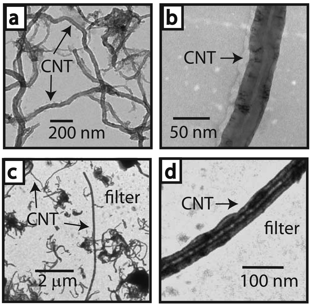
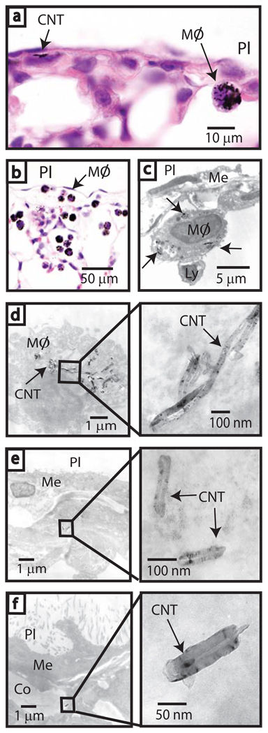
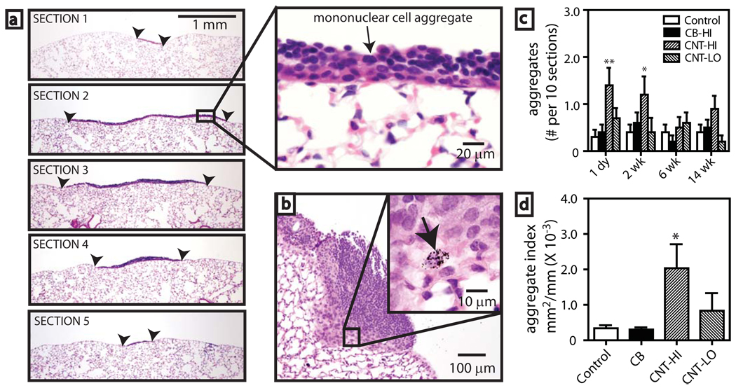
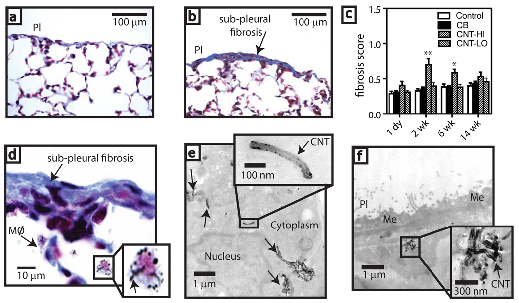
Carbon nanotubes are shaped like fibres and can stimulate inflammation at the surface of the peritoneum when injected into the abdominal cavity of mice, raising concerns that inhaled nanotubes may cause pleural fibrosis and/or mesothelioma. Here, we show that multiwalled carbon nanotubes reach the subpleura in mice after a single inhalation exposure of 30 mg m(-3) for 6 h. Nanotubes were embedded in the subpleural wall and within subpleural macrophages. Mononuclear cell aggregates on the pleural surface increased in number and size after 1 day and nanotube-containing macrophages were observed within these foci. Subpleural fibrosis unique to this form of nanotubes increased after 2 and 6 weeks following inhalation. None of these effects was seen in mice that inhaled carbon black nanoparticles or a lower dose of nanotubes (1 mg m(-3)). This work suggests that minimizing inhalation of nanotubes during handling is prudent until further long-term assessments are conducted.


Figures

Figure 1
Figure 1. Aerosolization of carbon nanotubes
a, Transmission electron micrograph (TEM) of bulk MWCNT prior to aerosolization. b, Higher magnification of an individual CNT in the bulk sample. c. Aerosolized CNT captured by electrostatic precipitation on a filter located within the inhalation tower port (see supplementary information). d. Higher magnification of an aerosolized precipitated CNT on filter.
Figure 2
Figure 2. Inhaled carbon nanotubes reach the sub-pleura in mice
a, Light microscopy of CNTs in sub-pleura and macrophage (MØ) indicated by arrows (hematoxylin and eosin, 1000X). b, Sub-pleural macrophages (MØ) with CNT 1 day after high dose inhalation (hematoxylin and eosin, 200X). c, TEM showing CNTs within a macrophage (arrows) beneath the pleura (PL). Relative location of the mesothelium (Me) and a lymphocyte (Ly) are indicated. d, TEM of CNT within a sub-pleural macrophage (inset shows detail). e, TEM of CNTs in a sub-pleural cell (inset shows detail). f, CNT in collagen (Co) beneath the mesothelium (Me) (see inset for detail).
Figure 3
Figure 3. Pleural immune response after carbon nanotube inhalation in mice
a, Sequential sections 100 µm apart through a pleural mononuclear cell aggregate (bordered by arrowheads) 1 day post-CNT inhalation (30 mg/m3) (hematoxylin and eosin, 40X). Inset shows detail (400X). b, Mononuclear aggregate 2 wk after CNT exposure (100X). Inset shows a macrophage with CNT indicated by arrow (1000X). c, Numbers of pleural aggregates after exposure to saline aerosol (control), 30 mg/m3 carbon black nanoparticles (CB), 30 mg/m3 CNT-(HI), or 1 mg/m3 CNT-(LO). Data are aggregates per 10 sections from each lung (N=10 animals). *P<0.05 or **P<0.001 compared to control. d, Aggregate size index measured by image analysis (see supplementary information). *P<0.05 compared to control determined by one-way ANOVA with post-hoc Tukey test.
Figure 4
Figure 4. Sub-pleural fibrosis in mice after carbon nanotube inhalation
Microscopy of trichrome-stained lung from a, saline aerosol-exposed (control) and b, CNT-exposed (30 mg/m3) mice 2 wk after inhalation (200X). c, Fibrosis score derived from a point-counting method after exposure to saline aerosol (control), 30 mg/m3 carbon black nanoparticles (CB), 30 mg/m3 CNT-(HI), or 1 mg/m3 CNT-(LO). *P<0.05 or **P<0.001 compared to control, CB, or CNT-LO determined by ANOVA with post-hoc Tukey test. d, Sub-pleural fibrotic lesion showing macrophages with CNT (inset shows detail). e, TEM of CNT within sub-pleural macrophage (arrows) at 2 wk. (Inset shows detail). f, TEM of CNT agglomerate in a sub-pleural cell at 6 wk (inset shows detail).

Comment in

  • Nanotoxicology: new insights into nanotubes.
    Donaldson K, Poland CA. Donaldson K, et al. Nat Nanotechnol. 2009 Nov;4(11):708-10. doi: 10.1038/nnano.2009.327. Epub 2009 Oct 25. Nat Nanotechnol. 2009. PMID: 19893519 No abstract available.

References

    1. Donaldson K, et al. Carbon nanotubes: a review of their properties in relation to pulmonary toxicology and workplace safety. Toxicol. Sci. 2006;92:5–22. - PubMed
    1. Poland CA, et al. Carbon nanotubes introduced into the abdominal cavity of mice show asbestos-like pathogenicity in a pilot study. Nat. Nanotech. 2008;3:423–428. - PubMed
    1. Ryman-Rasmussen JP, et al. Inhaled multiwalled carbon nanotubes potentiate airway fibrosis in a murine model of allergic asthma. Am. J. Resp. Cell. Mol. Biol. 2009;40:349–358. - PMC - PubMed
    1. Mossman BT, Churg A. Mechanisms in the pathogenesis of asbestosis and silicosis. Am. J. Resp. Crit. Care. Med. 1998;157:1666–1680. - PubMed
    1. Card JW, Zeldin DC, Bonner JC, Nestman ER. Pulmonary applications and toxicity of engineered nanoparticles. Am. J. Physiol. 2008;295:L400–L411. - PMC - PubMed

Publication types