. 2009 Nov;4(11):773-80.
doi: 10.1038/nnano.2009.294. Epub 2009 Oct 11.

A route to brightly fluorescent carbon nanotubes for near-infrared imaging in mice

Affiliations

A route to brightly fluorescent carbon nanotubes for near-infrared imaging in mice

Kevin Welsher et al. Nat Nanotechnol. 2009 Nov.

Abstract

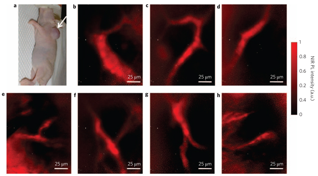
The near-infrared photoluminescence intrinsic to semiconducting single-walled carbon nanotubes is ideal for biological imaging owing to the low autofluorescence and deep tissue penetration in the near-infrared region beyond 1 microm. However, biocompatible single-walled carbon nanotubes with high quantum yield have been elusive. Here, we show that sonicating single-walled carbon nanotubes with sodium cholate, followed by surfactant exchange to form phospholipid-polyethylene glycol coated nanotubes, produces in vivo imaging agents that are both bright and biocompatible. The exchange procedure is better than directly sonicating the tubes with the phospholipid-polyethylene glycol, because it results in less damage to the nanotubes and improves the quantum yield. We show whole-animal in vivo imaging using an InGaAs camera in the 1-1.7 microm spectral range by detecting the intrinsic near-infrared photoluminescence of the 'exchange' single-walled carbon nanotubes at a low dose (17 mg l(-1) injected dose). The deep tissue penetration and low autofluorescence background allowed high-resolution intravital microscopy imaging of tumour vessels beneath thick skin.


Figures

Figure 1
Figure 1. Comparison of exchange- and direct-SWNTs
a, Schematic of the exchange process. Cholate (red and white balls) on SWNTs (grey) is dialysed and eventually replaced by phospholipid–polyethylene glycol (PL–PEG) to form biocompatible nanotubes without damaging the integrity of the nanotube sidewall. b, NIR photoluminescence images of the three solutions excited at 808 nm at equal concentrations. Exchange-SWNTs show greater fluorescence yield than direct-SWNTs. c, Photoluminescence versus excitation spectra show improved quantum yield in cholate and exchange samples. The dotted lines show how peaks are redshifted after exchange. d, UV–vis–NIR curves. Exchange- and cholate-SWNTs show sharp transition peaks; direct-SWNTs show very low and broad absorption features. PL, photoluminescence.
Figure 2
Figure 2. Atomic force microscopy and Raman spectroscopy
a,b, AFM topography images of exchange- (a) and direct- (b) SWNTs. The average lengths of the exchange and direct samples are 372 and 161 nm, respectively. c, Histogram of length distributions of exchange and direct samples. The exchange sample has lengths ranging from 50 nm up to 1.8 µm. The direct sample has a narrower distribution, with most SWNTs near 100 nm in length and none longer than 500 nm. d, Raman spectra of the three samples. The higher D band in direct-SWNTs indicates a greater number of defects and explains the lower quantum yield.
Figure 3
Figure 3. In vitro NIR photoluminescence targeted cell imaging
a–d, NIR photoluminescence images (1,100–1,700 nm) of human malignant glioma cells (U87 MG) treated with exchange-SWNT/RGD (a,b) and direct-SWNT/RGD (c,d) conjugates. Arginine–glycine–aspartic acid (RGD) peptide ligand binds to αvβ3-integrin overexpressed on the cells. Human breast cancer cell line (MDA-MB-468) is used as a negative. Cells treated with exchange-SWNT/RGD show a high positive signal with very low non-specific binding to the negative cell line. The direct-SWNT/RGD conjugates gave images with very little NIR photoluminescence signal.
Figure 4
Figure 4. In vivo NIR photoluminescence imaging of mice
a–f, NIR photoluminescence images (1,100–1,700 nm) of nude mice treated with 200 µl of 17 mg l−1 exchange-SWNTs (a–c) and 200µl of 260mg l−1 direct-SWNTs (d–f). Solution images show the difference in concentration of the injected doses of the exchange-SWNTs (inset, a) and direct-SWNTs (inset, d). Exchange-SWNTs show high image contrast despite their dose being a factor of 15 lower than that of the direct-SWNTs. Images were taken 30 min (b,e) and 24 h (c,f) post tail-vein injection. At early time points, the vasculature beneath the skin is visible as a result of the SWNTs circulating in the blood. The photoluminescence signal from deeper in the mouse is probably due to strong reticuloendothelial system uptake. g, NIR photoluminescence image of an untreated nude mouse showing ultralow autofluorescence. The photoluminescence signal from the SWNTs is easily distinguishable from the endogenous autofluorescence without any image processing. All images used an exposure time of 100 ms.
Figure 5
Figure 5. Intravital NIR photoluminescence imaging of tumour vessels
a, Optical image of LS174T tumour-bearing mouse used for intravital microscopy. b–h, High-magnification NIR photoluminescence images taken within 90 min of injection of a high concentration (~170 mg l−1, 300µl) of exchange-SWNTs. Tumour vessels can be resolved to a few micrometres, approaching the diffraction limit. Exposure times for all images were 1 s.

Comment in

References

    1. Liu Z, Tabakman S, Welsher K, Dai H. Carbon nanotubes in biology and medicine: in vitro and in vivo detection, imaging and drug delivery. Nano Res. 2009;2:85–175. - PMC - PubMed
    1. Dhar S, et al. Targeted single-wall carbon nanotube-mediated Pt(IV) prodrug delivery using folate as a homing device. J. Am. Chem. Soc. 2008;130:11467–11476. - PMC - PubMed
    1. Liu Z, Winters M, Holodniy M, Dai HJ. siRNA delivery into human T cells and primary cells with carbon-nanotube transporters. Angew Chem. Int. Ed. 2007;46:2023–2027. - PubMed
    1. Liu Z, et al. Drug delivery with carbon nanotubes for in vivo cancer treatment. Cancer Res. 2008;68:6652–6660. - PMC - PubMed
    1. Liu Z, et al. In vivo biodistribution and highly efficient tumour targeting of carbon nanotubes in mice. Nature Nanotech. 2007;2:47–52. - PubMed

Publication types

Substances