. 2010 Oct;113(4):826-34.
doi: 10.3171/2009.9.JNS09802.

Adenosine A(2A) receptors in early ischemic vascular injury after subarachnoid hemorrhage. Laboratory investigation

Affiliations

Adenosine A(2A) receptors in early ischemic vascular injury after subarachnoid hemorrhage. Laboratory investigation

Fatima A Sehba et al. J Neurosurg. 2010 Oct.

Abstract

Object: The role of adenosine A(2A) receptors in the early vascular response after subarachnoid hemorrhage (SAH) is unknown. In other forms of cerebral ischemia both activation and inhibition of A(2A) receptors is reported to be beneficial. However, these studies mainly used pharmacological receptor modulation, and most of the agents available exhibit low specificity. The authors used adenosine A(2A) receptor knockout mice to study the role of A(2A) receptors in the early vascular response to SAH.

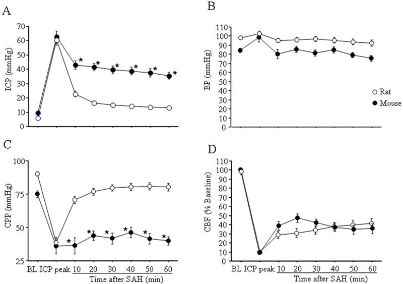
Methods: Subarachnoid hemorrhage was induced in wild-type mice (C57BL/6) and A(2A) receptor knockout mice by endovascular puncture. Cerebral blood flow, intracranial pressure, and blood pressure were recorded, and cerebral perfusion pressure was deduced. Animals were sacrificed at 1, 3, or 6 hours after SAH or sham surgery. Coronal brain sections were immunostained for Type IV collagen, the major protein of the basal lamina. The internal diameter of major cerebral arteries and the area fraction of Type IV collagen-positive microvessels (< 100 μm) were determined.

Results: The initial increase in intracranial pressure and decrease in cerebral perfusion pressure at SAH induction was similar in both types of mice, but cerebral blood flow decline was significantly smaller in A(2A) receptor knockout mice as compared with wild-type cohorts. The internal diameter of major cerebral vessels decreased progressively after SAH. The extent of diameter reduction was significantly less in A(2A) receptor knockout mice than in wild-type mice. Type IV collagen immunostaining decreased progressively after SAH. This decrease was significantly less in A(2A) receptor knockout mice than in wild-type mice.

Conclusions: These results demonstrate that global inactivation of A(2A) receptors decreases the intensity of the early vascular response to SAH. Early inhibition of A(2A) receptors after SAH might reduce cerebral injury.


Figures

Figure-1
Figure-1. A comparison of physiological changes after rat and mouse SAH induced via endovascular model
SAH rats that had similar ICP values as WT SAH mouse were used for this comparison. (Retrospective data from rat endovascular model) Note the difference in the pattern of ICP decline between the two species after SAH (A; see text for explanation). Baseline BP (B) was slightly lower in mouse and remained unchanged (as in rat) after SAH (B). Initial fall in CPP at SAH was the same but recovery was lower in mouse (C). CBF fall at SAH and recovery was the same between the two species (D).
Figure-2
Figure-2. A comparison of physiological changes after SAH in WT and A2AR-KO
Small but significant differences in physiological parameter after SAH were found. ICP rose to the similar value at SAH but decline to a smaller value at 10–20 minutes in A2AR-KO (A). At 60 minutes after SAH ICP plateau to the same value in WT and A2AR-KO mice. No difference in BP values before or after SAH occurred in WT and KO mice (B). Initial CPP fall was similar but a trend towards higher recovery thereafter was seen in A2AR-KO (C). CBF fell to a significantly smaller value in KO but recovered to the similar value at 60 minutes after SAH. * Significantly different than rat SAH at P<0.05.
Figure-3
Figure-3. Internal circumference of Major cerebral vessels in WT and A2AR-KO after SAH
A: Representative images of collagen-IV immunostained ICA from WT mice sacrificed 1 or 6 hours after sham or SAH surgeries. Internal circumference and wall thickness of 1hr-sham: 368μm and 41μm, 1hr-SAH: 191 and 66μm, 6hr-Sham: 305, and 39μm and, 6hr-SAH: 219 and 46μm. Scale bar: 50μm. B: Mice were sacrificed 1–6 hours after SAH or sham surgery. Internal circumference of WT shams was similar at 1 and 3 hours but decreased by 15% at 6 hours. Internal circumference A2AR-KO shams was similar to their time matched WT sham cohorts. In contrast, a significant time dependent reduction in internal circumference of WT and A2AR KO mice occurred after SAH. The magnitude of internal circumference reduction at 1 and 3hrs after SAH was significantly more in WT than in A2AR-KO. * Significantly different than sham at P<0.05.! Significantly different than A2AR-KO at P<0.05.
Figure-4
Figure-4. Collagen-IV immunostaining of parenchymal microvessels is reduced after SAH
A: Representative images of collagen-IV immunostaining of microvessels from striatum of WT and A2AR-KO mice sacrificed 1 hr after SAH. Note more complete and longer collagen-IV stained vascular profiles in A2AR-KO mice as compared to the time matched WT cohorts. B: Accumulative graph of the area fraction % respective sham of collagen-IV positive vascular profiles in WT and A2AR-KO mice sacrificed 1 to 6 hours after SAH. Note the delayed decrease in collagen-IV immunostaining in A2AR-KO as compared to WT mice after SAH. * Significantly different than sham operated animals at P<0.05,! Significantly different than the time matched A2AR-KO cohort at P<0.05.

References

    1. Aden U, Halldner L, Lagercrantz H, Dalmau I, Ledent C, Fredholm BB. Aggravated brain damage after hypoxic ischemia in immature adenosine A2A knockout mice. Stroke. 2003;34:739–744. - PubMed
    1. Asano T, Sano K. Pathogenetic role of no-reflow phenomenon in experimental subarachnoid hemorrhage in dogs. J Neurosurg. 1977;46:454–466. - PubMed
    1. Ayata C, Dunn AK, Gursoy OY, Huang Z, Boas DA, Moskowitz MA. Laser speckle flowmetry for the study of cerebrovascular physiology in normal and ischemic mouse cortex. J Cereb Blood Flow Metab. 2004;24:744–755. - PubMed
    1. Bederson JB, Germano IM, Guarino L. Cortical blood flow and cerebral perfusion pressure in a new noncraniotomy model of subarachnoid hemorrhage in the rat. Stroke. 1995;26:1086–1091. - PubMed
    1. Bederson JB, Levy AL, Ding WH, Kahn R, DiPerna CA, Jenkins ALr, et al. Acute vasoconstriction after subarachnoid hemorrhage. Neurosurgery. 1998;42:352–360. - PubMed

Publication types

MeSH terms

LinkOut - more resources