Early processing in the human lateral occipital complex is highly responsive to illusory contours but not to salient regions
- PMID: 19895562
- PMCID: PMC3224794
- DOI: 10.1111/j.1460-9568.2009.06981.x
Early processing in the human lateral occipital complex is highly responsive to illusory contours but not to salient regions
Abstract
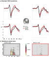
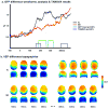
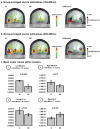
Human electrophysiological studies support a model whereby sensitivity to so-called illusory contour stimuli is first seen within the lateral occipital complex. A challenge to this model posits that the lateral occipital complex is a general site for crude region-based segmentation, based on findings of equivalent hemodynamic activations in the lateral occipital complex to illusory contour and so-called salient region stimuli, a stimulus class that lacks the classic bounding contours of illusory contours. Using high-density electrical mapping of visual evoked potentials, we show that early lateral occipital cortex activity is substantially stronger to illusory contour than to salient region stimuli, whereas later lateral occipital complex activity is stronger to salient region than to illusory contour stimuli. Our results suggest that equivalent hemodynamic activity to illusory contour and salient region stimuli probably reflects temporally integrated responses, a result of the poor temporal resolution of hemodynamic imaging. The temporal precision of visual evoked potentials is critical for establishing viable models of completion processes and visual scene analysis. We propose that crude spatial segmentation analyses, which are insensitive to illusory contours, occur first within dorsal visual regions, not the lateral occipital complex, and that initial illusory contour sensitivity is a function of the lateral occipital complex.
Figures


Similar articles
-
The Poggendorff illusion driven by real and illusory contour: Behavioral and neural mechanisms.Neuropsychologia. 2016 May;85:24-34. doi: 10.1016/j.neuropsychologia.2016.03.005. Epub 2016 Mar 5. Neuropsychologia. 2016. PMID: 26956926
-
Activation time course of responses to illusory contours and salient region: a high-density electrical mapping comparison.Brain Res. 2006 Feb 3;1071(1):137-44. doi: 10.1016/j.brainres.2005.11.089. Epub 2006 Jan 17. Brain Res. 2006. PMID: 16413507
-
Functional anatomy and differential time courses of neural processing for explicit, inferred, and illusory contours. An event-related fMRI study.Neuroimage. 2003 Aug;19(4):1567-77. doi: 10.1016/s1053-8119(03)00180-0. Neuroimage. 2003. PMID: 12948712
-
The representation of illusory and real contours in human cortical visual areas revealed by functional magnetic resonance imaging.J Neurosci. 1999 Oct 1;19(19):8560-72. doi: 10.1523/JNEUROSCI.19-19-08560.1999. J Neurosci. 1999. PMID: 10493756 Free PMC article.
-
fMRI activation in response to illusory contours and salient regions in the human lateral occipital complex.Neuron. 2003 Jan 23;37(2):323-31. doi: 10.1016/s0896-6273(02)01148-0. Neuron. 2003. PMID: 12546826 Clinical Trial.
Cited by
-
Feedback from lateral occipital cortex to V1/V2 triggers object completion: Evidence from functional magnetic resonance imaging and dynamic causal modeling.Hum Brain Mapp. 2021 Dec 1;42(17):5581-5594. doi: 10.1002/hbm.25637. Epub 2021 Aug 21. Hum Brain Mapp. 2021. PMID: 34418200 Free PMC article.
-
Early electrophysiological indices of illusory contour processing within the lateral occipital complex are virtually impervious to manipulations of illusion strength.Neuroimage. 2012 Feb 15;59(4):4074-85. doi: 10.1016/j.neuroimage.2011.10.051. Epub 2011 Oct 21. Neuroimage. 2012. PMID: 22037001 Free PMC article.
-
The strength of feedback processing is associated with resistance to visual backward masking during Illusory Contour processing in adult humans.Neuroimage. 2022 Oct 1;259:119416. doi: 10.1016/j.neuroimage.2022.119416. Epub 2022 Jun 25. Neuroimage. 2022. PMID: 35764208 Free PMC article.
-
The effort to close the gap: tracking the development of illusory contour processing from childhood to adulthood with high-density electrical mapping.Neuroimage. 2014 Apr 15;90:360-73. doi: 10.1016/j.neuroimage.2013.12.029. Epub 2013 Dec 21. Neuroimage. 2014. PMID: 24365674 Free PMC article.
-
Brain network mechanisms of visual shape completion.Neuroimage. 2021 Aug 1;236:118069. doi: 10.1016/j.neuroimage.2021.118069. Epub 2021 Apr 18. Neuroimage. 2021. PMID: 33878383 Free PMC article.
References
-
- Anderson BL. Filling-in models of completion: rejoinder to Kellman, Garrigan, Shipley, and Keane (2007) and Albert (2007) Psychol Rev. 2007;114(2):509–27. - PubMed
-
- Bar M. A cortical mechanism for triggering top-down facilitation in visual object recognition. J Cogn Neurosci. 2003;15:600–609. - PubMed
-
- Chen CM, Lakatos P, Shah AS, Mehta AD, Givre SJ, Javitt DC, Schroeder CE. Functional Anatomy and Interaction of Fast and Slow Visual Pathways in Macaque Monkeys. Cereb Cortex 2006 - PubMed
Publication types
MeSH terms
Substances
Grants and funding
LinkOut - more resources
Full Text Sources

