. 2010 Jan;112(2):434-43.
doi: 10.1111/j.1471-4159.2009.06477.x. Epub 2009 Nov 6.

Synergistic dopaminergic neurotoxicity of manganese and lipopolysaccharide: differential involvement of microglia and astroglia

Affiliations

Synergistic dopaminergic neurotoxicity of manganese and lipopolysaccharide: differential involvement of microglia and astroglia

Ping Zhang et al. J Neurochem. 2010 Jan.

Abstract

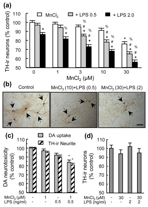
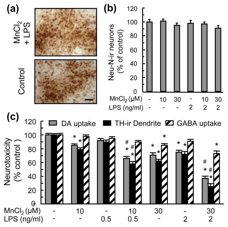
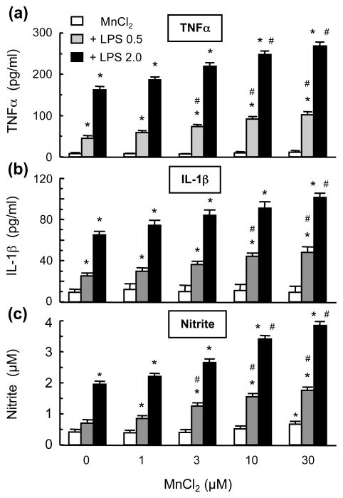
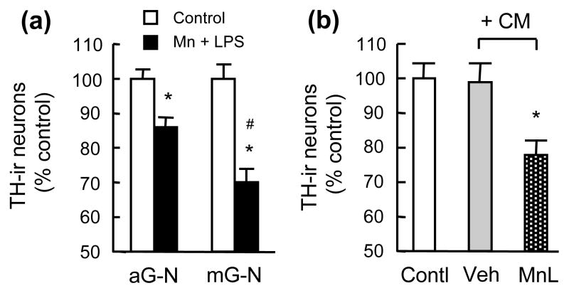
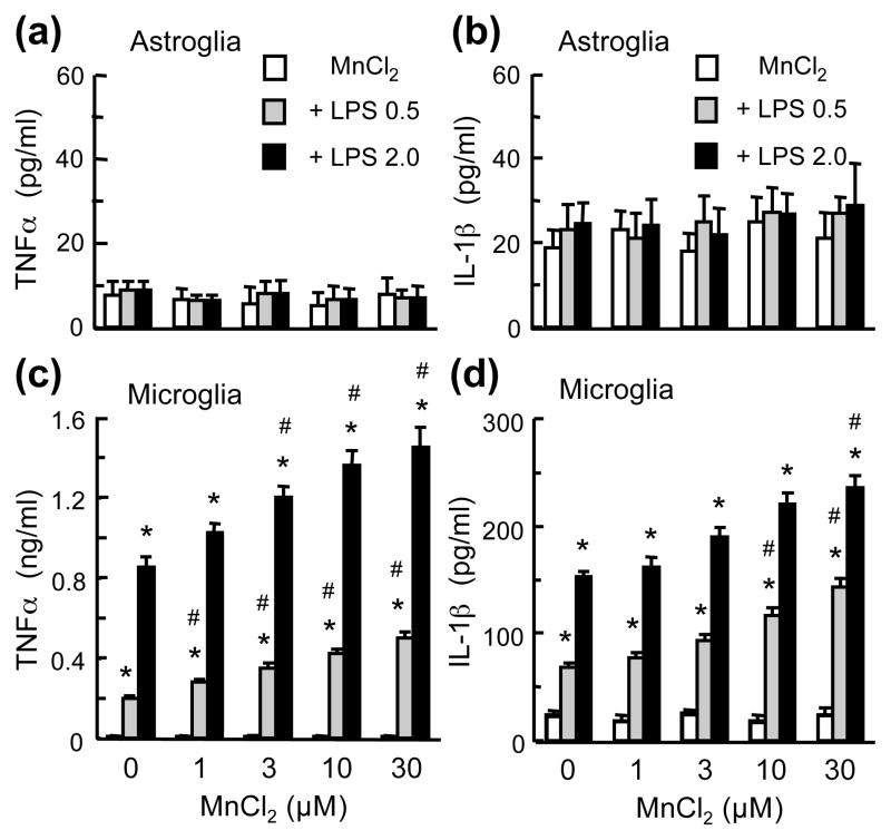
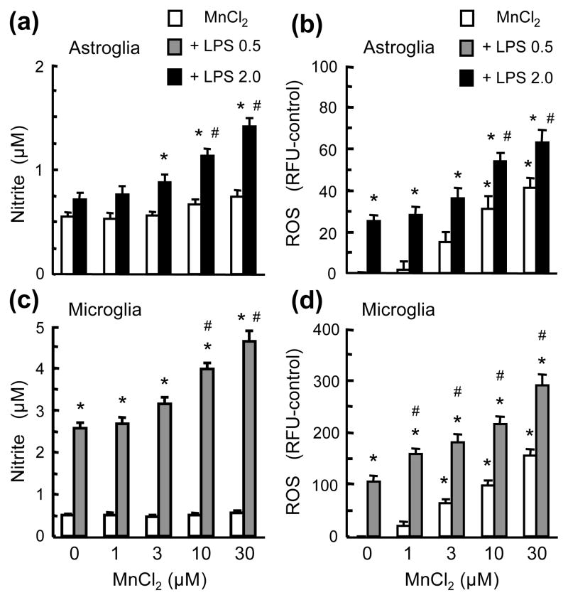
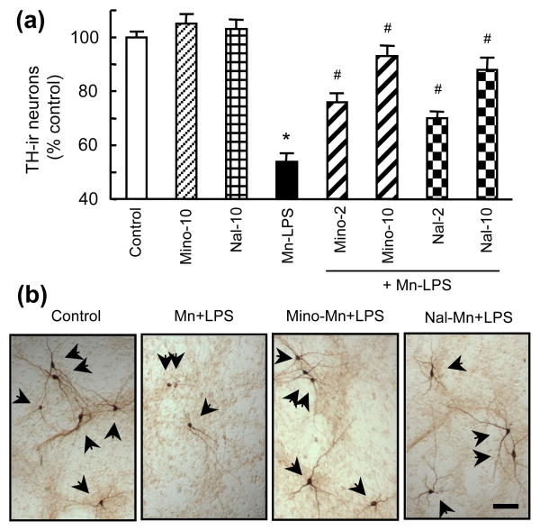
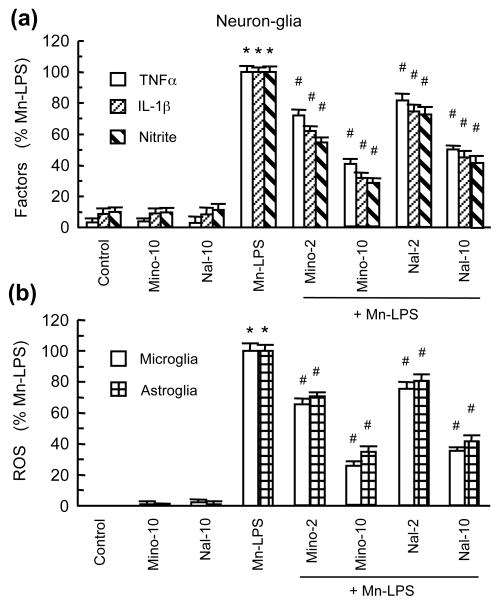
Overexposure to manganese is known to cause damage to basal ganglial neurons and the development of movement abnormalities. Activation of microglia and astrocytes has increasingly been associated with the pathogenesis of a variety of neurological disorders. We have recently shown that microglial activation facilitates manganese chloride (MnCl2, 10-300 microM)-induced preferential degeneration of dopamine (DA) neurons. In this study, we report that combinations of MnCl2 (1-30 microM) and endotoxin lipopolysaccharide (LPS, 0.5-2 ng/mL), at minimally effective concentrations when used alone, induced synergistic and preferential damage to DA neurons in rat primary neuron-glia cultures. Mechanistically, MnCl2 significantly potentiated LPS-induced release of tumor necrosis factor-alpha and interleukin-1 beta in microglia, but not in astroglia. MnCl2 and LPS were more effective in inducing the formation of reactive oxygen species and nitric oxide in microglia than in astroglia. Furthermore, MnCl2 and LPS-induced free radical generation, cytokine release, and DA neurotoxicity was significantly attenuated by pre-treatment with potential anti-inflammatory agents minocycline and naloxone. These results demonstrate that the combination of manganese overexposure and neuroinflammation is preferentially deleterious to DA neurons. Moreover, these findings not only shed light on the understanding of manganese neurotoxicity but may also bear relevance to the potentially multifactorial etiology of Parkinson's disease.


Figures

Fig. 1
Fig. 1
DA neurotoxicity induced by combined treatment with MnCl2 and LPS. Rat primary mesencephalic neuron-glia cultures (a–c) or neuron-enriched cultures (d) were treated for 7 days with vehicle control (0.2% water), indicated concentrations of MnCl2 or LPS alone, and combinations of MnCl2 and LPS. Following treatment, the cultures were either immunostained for TH-ir neurons (a–d) or subject to measurement of [3H]DA uptake capacity (c). The number of TH-ir neurons (a & d) and neurite length of TH-ir neurons (c) were determined. Results are expressed as a percentage of the vehicle-treated control cultures and are mean ± SEM of three (d) or five (a & c) independent experiments performed in triplicate. *p < 0.05 compared to vehicle-treated control cultures, #p < 0.05 compared to corresponding LPS-treated cultures, %p < 0.05 compared to corresponding MnCl2-treated cultures. In (b), the numbers in parentheses of indicate 10 or 30 μM and 0.5 or 2 ng/ml respectively. ICC images (b) are from a representative experiment. Arrowheads: TH-ir neurons. Scale bar: 100 μm.
Fig. 2
Fig. 2
Preferential DA neurotoxicity induced by MnCl2 and LPS. Neuron-glia cultures were treated for 7 days with vehicle, MnCl2 or LPS alone, and combinations of MnCl2 and LPS. Cultures were then immunostained for counting Neu-N-ir neurons (b) and measurement of TH-ir neurites (c), or assayed for [3H]DA and [3H]GABA uptake (c). For ICC analysis (a), cultures were treated with vehicle (Control) or 30 μM MnCl2 and 2 ng/ml LPS (MnCl2 + LPS), scale bar: 50 μm. Results are expressed as a percentage of the vehicle-treated control cultures and are mean ± SEM of four (b) or five (c) experiments performed in triplicate. *p< 0.05 compared to vehicle-treated control cultures, #p < 0.05 compared to corresponding MnCl2 or LPS-treated cultures.
Fig. 3
Fig. 3
Effect of MnCl2 and LPS on the release of TNFα and IL-1β and production of NO in neuron-glia cultures. Supernatants from cultures treated for 7 days with vehicle, MnCl2 or LPS alone, and combinations of MnCl2 and LPS were collected and the levels of TNFα, IL-1β, and nitrite were determined. Results are mean ± SEM of five experiments performed in triplicate. *p< 0.05 compared to vehicle-treated control cultures, #p < 0.05 compared to corresponding MnCl2 or LPS-treated cultures.
Fig. 4
Fig. 4
Characterization of glial dependence for MnCl2 and LPS-induced DA neurotoxicity. (a). Astroglia-neuron (aG-N) or microglia-neuron (mG-N) cultures were treated for 7 days with vehicle and 30 μM MnCl2 and 2 ng/ml LPS (Mn+LPS). (b). Neuron-enriched cultures were treated for 6 days with treatment media alone (Contl) or treatment media containing conditioned media (CM) collected from microglia treated for 24 hr with vehicle (Veh) or 30 μM MnCl2 and 2 ng/ml LPS (MnL). Following treatment, cultures were immunostained and the number of TH-ir neurons determined. Results are mean ± SEM of four experiments performed in triplicate. *p< 0.05 compared to vehicle-treated control cultures, #p < 0.05 compared to MnCl2+LPS-treated astroglia-neuron cultures.
Fig. 5
Fig. 5
Comparison of the effect of MnCl2 and LPS on TNFα and IL-1β release in microglia and astroglia. Astroglia (a & b) microglia (c & d) cultures were treated for 24 hr with vehicle, MnCl2 or LPS alone, and the combinations of MnCl2 and LPS. The levels of TNFα (a & c) and IL-1β (b & d) in the culture supernatants were determined. Results are mean ± SEM of four to six experiments performed in triplicate. *p< 0.05 compared to vehicle-treated control cultures, #p < 0.05 compared to corresponding MnCl2 or LPS-treated cultures.
Fig. 6
Fig. 6
Effect of MnCl2 and LPS on NO and ROS production in microglia and astroglia. Astroglia (a & b) microglia (c & d) cultures were treated for 24 hr (b–d) or 72 hr (a) with vehicle, MnCl2 or LPS alone, and the combinations of MnCl2 and LPS and production of NO and ROS was determined. Results are mean ± SEM of four to six experiments performed in triplicate. *p< 0.05 compared to vehicle-treated control cultures, #p < 0.05 compared to corresponding MnCl2 or LPS-treated cultures.
Fig. 7
Fig. 7
Effect of naloxone and minocycline on the MnCl2 and LPS-induced DA neurodegeneration. Neuron-glia cultures were treated for 7 days with vehicle, 10 μM minocycline (Mino-10) or naloxone (Nal-10) alone, the combination of 30 μM MnCl2 and 2 ng/ml LPS (Mn-LPS), or pretreated for 30 min with 2 or 10 μM minocycline (Mino-2, Mino-10) or naloxone (Nal-2, Nal-10) prior to treatment with 30 μM MnCl2 and 2 ng/ml LPS (+ Mn-LPS). Cultures were then immunostained for TH-ir neurons and the number of TH-ir neurons was determined. Results in (a) are mean ± SEM of five experiments performed in triplicate. *p< 0.05 compared to vehicle-treated control cultures, #p < 0.05 compared to MnCl2 and LPS-treated cultures. ICC images (b) are from cultures treated with vehicle (control), 30 μM MnCl2 and 2 ng/ml LPS (Mn+LPS), and pretreated with 10 μM minocycline or naloxone prior to treatment with MnCl2 and LPS (Mino-Mn+LPS or Nal-Mn+LPS). Bar: 100 μm.
Fig. 8
Fig. 8
Effect of minocycline and naloxone on the MnCl2 and LPS-induced production of neurotoxic factors. (a). The levels of TNFα, IL-1β, and nitrite in the supernatants from cultures treated as described for Fig. 7a were determined. (b). Microglia and astroglia cultures were treated for 24 hr with the same combinations of agents as described for Fig. 7a and ROS production was determined. Results in are mean ± SEM of five (a) or four (b) experiments performed in triplicate (a) or quadruplicate (b). *p< 0.05 compared to vehicle-treated control cultures, #p < 0.05 compared to MnCl2 and LPS-treated cultures.

References

    1. Aschner M, Guilarte TsR, Schneider JS, Zheng W. Manganese: Recent advances in understanding its transport and neurotoxicity. Toxicology and applied pharmacology. 2007;221:131–147. - PMC - PubMed
    1. Bae JH, Jang BC, Suh SI, Ha E, Baik HH, Kim SS, Lee MY, Shin DH. Manganese induces inducible nitric oxide synthase (iNOS) expression via activation of both MAP kinase and PI3K/Akt pathways in BV2 microglial cells. Neuroscience letters. 2006;398:151–154. - PubMed
    1. Barhoumi R, Faske J, Liu X, Tjalkens RB. Manganese potentiates lipopolysaccharide-induced expression of NOS2 in C6 glioma cells through mitochondrial-dependent activation of nuclear factor kappaB. Brain Res Mol Brain Res. 2004;122:167–179. - PubMed
    1. Brydon L, Harrison NA, Walker C, Steptoe A, Critchley HD. Peripheral inflammation is associated with altered substantia nigra activity and psychomotor slowing in humans. Biol Psychiatry. 2008;63:1022–1029. - PMC - PubMed
    1. Calne DB, Chu NS, Huang CC, Lu CS, Olanow W. Manganism and idiopathic parkinsonism: similarities and differences. Neurology. 1994;44:1583–1586. - PubMed

Publication types

MeSH terms