. 2009 Nov 8:10:81.
doi: 10.1186/1471-2121-10-81.

The proline-rich domain of tau plays a role in interactions with actin

Affiliations

The proline-rich domain of tau plays a role in interactions with actin

Hai Jin He et al. BMC Cell Biol. .

Abstract

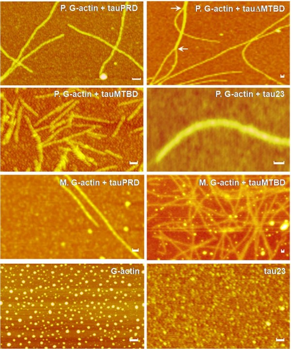
Background: The microtubule-associated protein tau is able to interact with actin and serves as a cross-linker between the microtubule and actin networks. The microtubule-binding domain of tau is known to be involved in its interaction with actin. Here, we address the question of whether the other domains of tau also interact with actin.

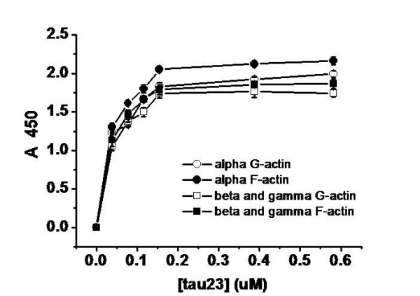
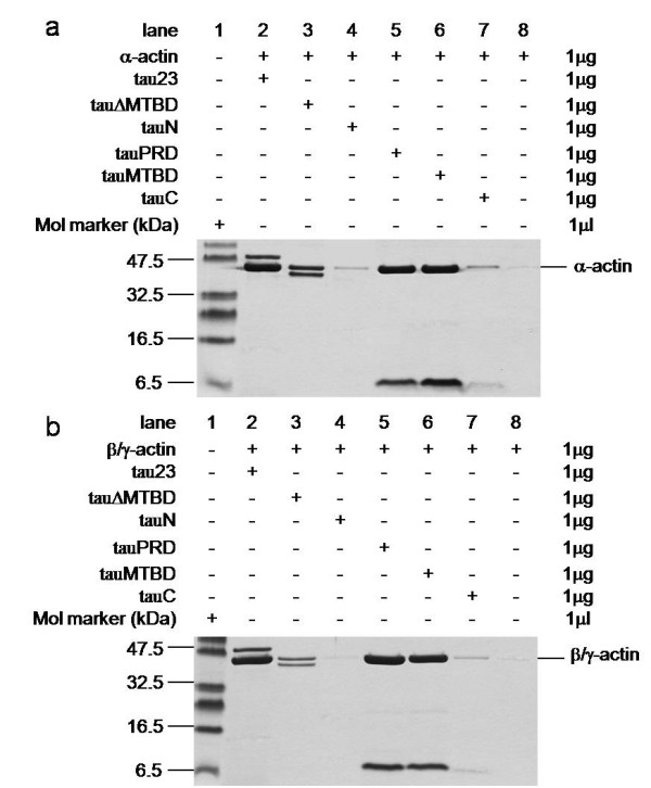
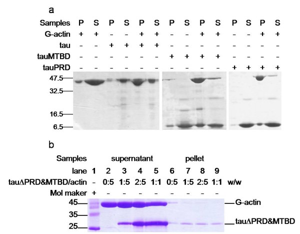
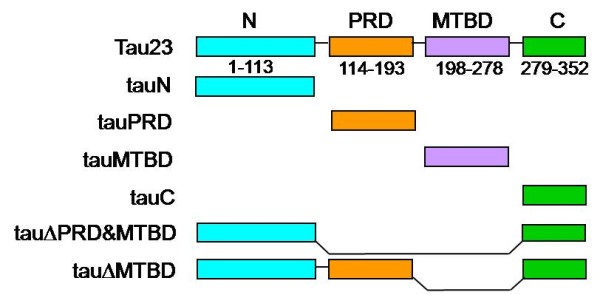
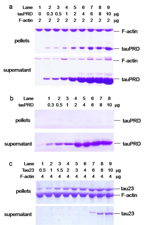
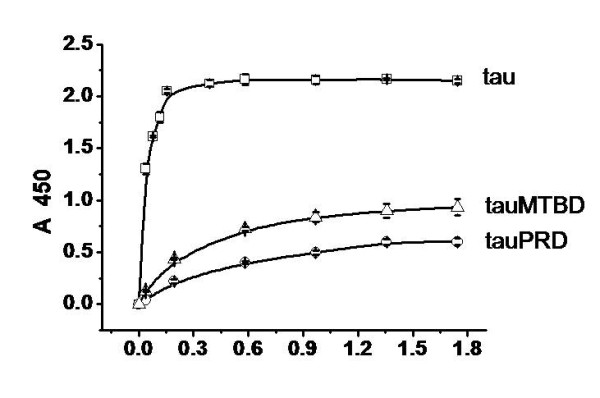
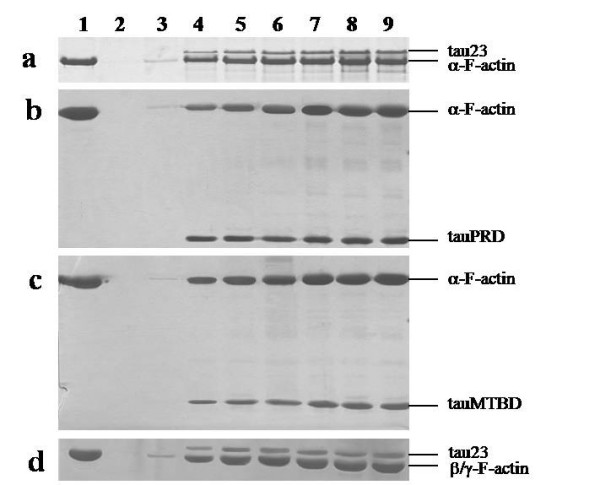
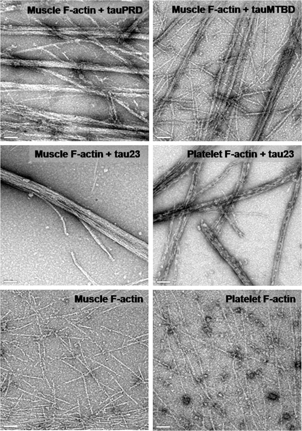
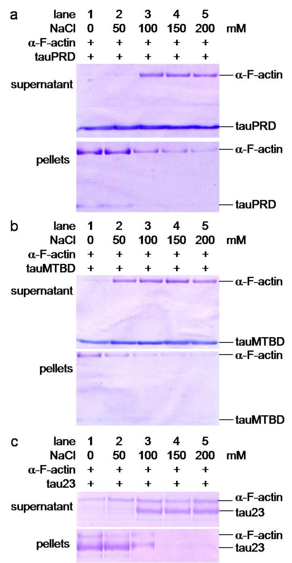
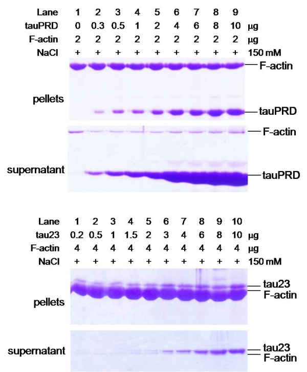
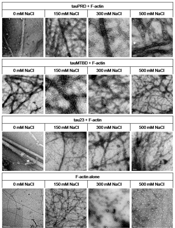
Results: Several tau truncation and deletion mutants were constructed, namely N-terminal region (tauN), proline-rich domain (tauPRD), microtubule binding domain (tauMTBD) and C-terminal region (tauC) truncation mutants, and microtubule binding domain (tauDeltaMTBD) and proline-rich domain/microtubule binding domain (tauDeltaPRD&MTBD) deletion mutants. The proline-rich domain truncation mutant (tauPRD) and the microtubule binding domain deletion mutant (tauDeltaMTBD) promoted the formation of actin filaments. However, actin assembly was not observed in the presence of the N-terminal and C-terminal truncation mutants. These results indicate that the proline-rich domain is involved in the association of tau with G-actin. Furthermore, results from co-sedimentation, solid phase assays and electron microscopy showed that the proline-rich domain is also capable of binding to F-actin and inducing F-actin bundles. Using solid phase assays to analyze apparent dissociation constants for the binding of tau and its mutants to F-actin resulted in a sequence of affinity for F-actin: tau >> microtubule binding domain > proline-rich domain. Moreover, we observed that the proline-rich domain was able to associate with and bundle F-actin at physiological ionic strength.

Conclusion: The proline-rich domain is a functional structure playing a role in the association of tau with actin. This suggests that the proline-rich domain and the microtubule-binding domain of tau are both involved in binding to and bundling F-actin.


Figures

Figure 1
Figure 1
Binding of tau to actin detected by solid phase assay. Wells were coated with 50 μl G-actin and F-actin (5 μg/ml) from skeletal muscle and platelets, respectively. Aliquots of different concentrations of tau (from 0.03 to 0.55 μM) were incubated with actin in binding buffer (50 mM Hepes containing 0.5 mM EGTA and 0.5 mM MgCl2, pH 7.5). Immunoreactivity was monitored in the solid phase assay using secondary antibody-labelled HRP. Each point represents the mean of five determinations.
Figure 2
Figure 2
Low speed co-sedimentation assay of G-actin in the presence of tau and tau mutants. Tau and tau mutants were incubated with G-actin in a 1:1 mass ratio at 37°C in binding buffer and then centrifuged (25,000 g, 4°C, 30 min). Before electrophoresis on gels, pellets were resuspended with 20 mM Tris-HCl (pH 8.0) and boiled for 5 min. Rabbit skeletal muscle G-actin (panel a) and human platelet G-actin (panel b) were used. Actin alone was used as negative control. Different concentrations of tauΔPRD&MTBD reacted with G-actin. The pellets were electrophoresed on 12% SDS-PAGE gels after the centrifugation (panel c).
Figure 3
Figure 3
G-actin in the presence of tau mutants at low speed co-sedimentation assay. Conditions were referred to Figure 2, G-actin (4 μg) was incubated with equal amount of tauRPD, tauMTBD, tau (panel a), and with tauΔPRD&MTBD (panel b) in binding buffer for 20 min in 37°C, followed by low-speed centrifugation. The pellet fraction (P) and supernatant fraction (S) were loaded in gels.
Figure 4
Figure 4
Construction of tau and its mutants. Six truncated tau isolation and deletion mutants were constructed: tauN (N-terminal region, 1-113), tauPRD (proline-rich domain, 114-193, containing 22 proline residues), tauMTBD (microtubule-binding domain, 198-278) and tauC (C-terminal region, 279-352). TauΔMTBD is a mutant in which the MTBD domain is deleted. TauΔPRD&MTBD is a mutant in which both PRD and MTBD are deleted.
Figure 5
Figure 5
Interactions of tauPRD with G-actin observed by atomic force microscopy. Skeletal muscle (M) and platelet (P) G-actin were incubated with tauPRD in binding buffer at 37°C for 30 min and then aliquots were taken for observation by atomic force microscopy as indicated. Bars in panels are 50 nm.
Figure 6
Figure 6
High speed co-sedimentation assay of tauPRD and F-actin. F-actin (at a constant concentration) was incubated with different concentrations of tauPRD (panel a), tauPRD alone (panel b) and tau (panel c) in the binding buffer without addition of extra NaCl at 37°C for 40 min and then centrifuged (100,000 g, 4°C, 60 min). The pellet and supernatant were electrophoresed on 12% SDS-PAGE gels.
Figure 7
Figure 7
Solid phase assay studies on the interaction of tauPRD with F-actin. 50 μl skeletal muscle F-actin (5 μg/ml) was added to solid phase assay wells. Aliquots of different concentrations of tauPRD (○), tau (□) and tauMTBD (△) were incubated with actin in binding buffer. Immunoreactivity was monitored using secondary antibody-labelled HRP. Each point represents the mean of five determinations. Binding parameters were estimated by fitting data using the Hyperbl function in Origin (Table 2).
Figure 8
Figure 8
Low speed co-sedimentation assay of F-actin in the presence of tauPRD. Conditions were the same as those described for Figure 4, except that F-actin (5 μg) from skeletal muscle (used in panels a, b and c) and platelet actin (used in panel d) were incubated with tau at concentrations of 0, 1, 3, 5, 7, 10, and 15 μM (lanes 3 to 9 in each panel) and with tauPRD or tauMTBD at concentrations of 0, 5, 10, 15, 25, 35, and 45 μM (lanes 3 to 9). After sedimentation at 25,000 g, pellets of bundles were electrophoresed on SDS-PAGE gels. Total amount of F-actin (lane 1) and BSA alone (lane 2) were used as controls.
Figure 9
Figure 9
Electron microscope images of F-actin incubated with tauPRD. F-actin was incubated with tauPRD, tauMTBD and tau23 as described in Figure 4 and visualized with electron microscope. F-actin alone was shown as negative control. Bars in panels are 100 nm.
Figure 10
Figure 10
Low speed Co-sedimentation assays of tauPRD in the presence of F-actin in the presence of NaCl. Conditions were the same as those in Figure 4, except that F-actin was used instead of G-actin. F-actin was incubated with tauPRD, tau or tauMTBD at different concentrations of NaCl, and precipitates were electrophoresed on SDS-PAGE gels as indicated.
Figure 11
Figure 11
High speed co-sedimentation assays of tauPRD and F-actin in the presence of NaCl. F-actin (at a constant amount) was incubated with different amounts of tauPRD (panel a) and tau (panel b) in binding buffer with 150 mM NaCl at 37°C for 40 min to mimic physiological ionic strength and then centrifuged (100,000 g, 4°C, 60 min). The pellet and supernatant were electrophoresed on 12% SDS-PAGE gels.
Figure 12
Figure 12
Electron microscopy images of F-actin incubated with tauPRD in the presence of NaCl at different concentrations. Conditions were the same as those in Figure 7, except that different concentrations of NaCl were added to the reaction mixtures of F-actin and tauPRD (tau or tauMTBD) as indicated. The bar in each panel represents 200 nm.

References

    1. Weingarten MD, Lockwood AH, Hwo SY, Kirschner MW. A protein factor essential for microtubule assembly. Proc Natl Acad Sci USA. 1975;72(5):1858–1862. doi: 10.1073/pnas.72.5.1858. - DOI - PMC - PubMed
    1. Drechsel DN, Hyman AA, Cobb MH, Kirschner MW. Modulation of the dynamic instability of tubulin assembly by the microtubule-associated protein tau. Mol Biol Cell. 1992;3(10):1141–1154. - PMC - PubMed
    1. Brandt R. The tau proteins in neuronal growth and development. Front Biosci. 1996;1:d118–130. - PubMed
    1. Henriquez JP, Cross D, Vial C, Maccioni RB. Subpopulations of tau interact with microtubules and actin filaments in various cell types. Cell Biochem Funct. 1995;13(4):239–250. doi: 10.1002/cbf.290130404. - DOI - PubMed
    1. Farias GA, Munoz JP, Garrido J, Maccioni RB. Tubulin, actin, and tau protein interactions and the study of their macromolecular assemblies. J Cell Biochem. 2002;85(2):315–324. doi: 10.1002/jcb.10133. - DOI - PubMed

Publication types

LinkOut - more resources