

Cardiomyocyte autophagy: remodeling, repairing, and reconstructing the heart
- PMID: 19895751
- PMCID: PMC3005716
- DOI: 10.1007/s11906-009-0070-1
Cardiomyocyte autophagy: remodeling, repairing, and reconstructing the heart
Abstract
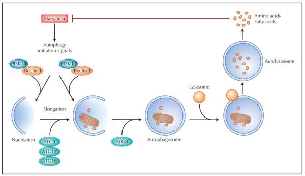
Autophagy is an evolutionarily conserved catabolic pathway of lysosome-dependent turnover of damaged proteins and organelles. When nutrients are in short supply, bulk removal of cytoplasmic components by autophagy replenishes depleted energy stores, a process critical for maintaining cellular homeostasis. However, prolonged activation of autophagic pathways can result in cell death. Longstanding evidence has linked the stimulation of lysosomal pathways to pathologic cardiac remodeling and a number of cardiac diseases, including heart failure and ischemia. Only recently, however, has work begun to parse cytoprotective autophagy from autophagy that contributes to disease pathogenesis. Current thinking suggests that the effects of autophagy exist on a continuum, with the eliciting triggers, the duration and amplitude of autophagic flux, and possibly the targeted intra-cellular cargo as critical determinants of the end result. Deciphering how autophagy participates in basal homeostasis of the heart, in aging, and in disease pathogenesis may uncover novel insights with clinical relevance in the treatment of heart disease.
Figures
References
-
- Lloyd-Jones D, Adams R, Carnethon M, et al. Heart disease and stroke statistics—2009 update: a report from the American Heart Association Statistics Committee and Stroke Statistics Subcommittee. Circulation. 2009;119:e21–e181. - PubMed
-
- Hill JA, Olson EN. Cardiac plasticity. N Engl J Med. 2008;358:1370–1380. - PubMed
-
- Nakatogawa H, Suzuki K, Kamada Y, Ohsumi Y. Dynamics and diversity in autophagy mechanisms: lessons from yeast. Nat Rev Mol Cell Biol. 2009;10:458–467. - PubMed
-
-
Levine B, Kroemer G. Autophagy in aging, disease and death: the true identity of a cell death impostor. Cell Death Differ. 2009;16:1–2. This review summarizes the major recent advances in autophagy in the contexts of disease and aging.
-
Publication types
MeSH terms
Grants and funding
LinkOut - more resources
Full Text Sources
Medical