Review
. 2009 Nov;7(11 Suppl):S68-78.
doi: 10.1016/j.cgh.2009.08.023.

Intrahepatic cholangiocarcinoma progression: prognostic factors and basic mechanisms

Affiliations
Review

Intrahepatic cholangiocarcinoma progression: prognostic factors and basic mechanisms

Alphonse E Sirica et al. Clin Gastroenterol Hepatol. 2009 Nov.

Abstract

In this review, we will examine various molecular biomarkers for their potential to serve as independent prognostic factors for predicting survival outcome in postoperative patients with progressive intrahepatic cholangiocarcinoma. Specific rodent models of intrahepatic cholangiocarcinoma that mimic relevant cellular, molecular, and clinical features of the human disease are also described, not only in terms of their usefulness in identifying molecular pathways and mechanisms linked to cholangiocarcinoma development and progression, but also for their potential value as preclinical platforms for suggesting and testing novel molecular strategies for cholangiocarcinoma therapy. Last, recent studies aimed at addressing the role of desmoplastic stroma in promoting intrahepatic cholangiocarcinoma progression are highlighted in an effort to underline the potential value of targeting tumor stromal components together with that of cholangiocarcinoma cells as a novel therapeutic option for this devastating cancer.


Conflict of interest statement

Conflicts of interest: The authors disclose no conflicts.

Figures

Figure 1
Figure 1
Differential expression of MUC1 and MUC2 in human intestinal-type (Int-CC) versus tubular-type (Tub-CC) human intrahepatic cholangiocarcinoma. (A) Low-grade papillary intestinal-type cholangiocarcinoma exhibiting an extensive goblet cell metaplasia. U, upper region of neoplastic papillae; L, lower or “cryptic” region of neoplastic papillae. (B) MUC2-positive neoplastic epithelial cells within the neoplastic papillae of a low-grade Int-CC. (C) Well-differentiated Tub-CC showing a prominent desmoplastic stroma. (D) Neoplastic glands of a Tub-CC exhibiting uniformly strong positive cell membrane and cytoplasmic immunoreactivity for MUC1. (E) Distribution of “intestinal” differentiation biomarkers (goblet cells and MUC2-positive cells) in Int-CC versus Tub-CC, compared with intrahepatic bile ducts of normal adult liver (BD), as well as with small (SD, ≤500 μm in diameter) and large (LD, ≥1000 μm in diameter) hyperplastic intrahepatic bile ducts in primary sclerosing cholangitis (PSC) liver. Note distribution of MUC2-positive cells closely parallels that of metaplastic goblet cells. (F) Comparison of levels of MUC1 immunostaining, reflected by mean optical density intensity (MOI) values, exhibited by malignant neoplastic epithelial cells of Tub-CC versus Int-CC (U and L) and relative to MOI values determined for non-neoplastic BD, PSC-SD, and PSC-LD, respectively. (G) Linear regression curve for MUC1 immunostaining intensity (MOI) values versus nuclear Ki-67 labeling indices in Tub-CC (f), Int-CC-L (e), and-U (d), and PSC-LD (c) PSC-SD (b), and BD (a). R, correlation coefficient.
Figure 2
Figure 2
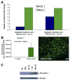
(A) Real-time RT-PCR gene expression measurements of amphiregulin (Areg) and mucin1 (Muc1) mRNA expressed in neoplastic biliary ducts obtained by laser capture microdissection from rat BDEneu intrahepatic cholangiocarcinomas at 10 and 25 days after inoculation. Glyceraldehyde 3-phosphate dehydrogenase–normalized fold changes are plotted. Note that amphiregulin mRNA is up-regulated in the malignant cholangiocytes in day 25 liver tumor compared with day 10 tumor, whereas MUC1 is equally expressed in both. (B) Representative photomicrograph depicting strongly positive immunofluorescence staining for caveolin-1 overexpressed in neoplastic cholangiocytes of a rat BDEneu intrahepatic cholangiocarcinoma, together with corresponding mean MOI values ± standard deviation for caveolin-1 immunostaining in tissue sections from day 25 BDEneu liver tumors (n = 3) compared with pair-matched cancer-free right liver lobe tissue samples obtained from the same animals as the tumor. Caveolin-1 immunoreactivity was not detected in either normal (day 10 liver) or hyperplastic bile ducts (day 25 liver) observed in the corresponding pair-matched rat liver lobe tissue samples. However, a strong positive immunoreactivity was observed in the portal vein and artery branches of the rat liver lobe tissue samples, as well as in the BDEneu tumor vasculature (data not shown). (C) Representative Western blot demonstrating prominently overexpressed caveolin-1 protein in whole tumor lysates prepared from day 25 rat BDEneu liver tumor (T 25) and associated peritoneal metastasis (Met 25) relative to caveolin-1 protein levels expressed in normal adult rat liver (NL), cholestatic liver at 21 days after bile duct ligation (BDL), and right liver lobe without cancer (RL) from the same rat as the BDEneu tumor.
Figure 3
Figure 3
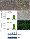
(A) Representative photomicrograph of rat BDEneu liver tumor tissue section exemplifying positive cytoplasmic immunoreactivity for “biliary” cytokeratin 19 as a characteristic phenotypic feature of neoplastic cholangiocytes in intrahepatic cholangiocarcinoma. (B) Photomicrograph depicting abundant α-SMA–positive intratumoral stromal cells surrounding cholangiocarcinoma cell nests (cc) in an invasive rat BDEneu liver tumor. P, portal area; H, hepatocytes. Representative photomicrographs depicting strong positive immunostaining reactions, together with corresponding MOI values (mean ± standard deviation), for the stromal proteins tenascin (C) and periostin (D) in tissue sections from day 25 BDEneu liver tumors compared with respective cancer-free pair-matched right liver lobe tissue samples. Tenascin and periostin immunostaining was either not detected or only marginally detected in the analyzed non-cancerous liver tissue sections with or without bile duct hyperplasia. (E) Representative Western blot demonstrating profound differential overexpression of periostin in day 25 rat BDEneu intrahepatic cholangiocarcinoma (T 25) and associated peritoneal metastatic tumor (Met 25) compared with normal adult rat liver (NL), 21 day bile duct–ligated liver (BDL), and pair-matched right liver lobe without cancer (RL 25).

References

    1. Hammill CW, Wong LL. Intrahepatic cholangiocarcinoma: a malignancy of increasing importance. J Am Coll Surg. 2008;207:594–603. - PubMed
    1. Khan SA, Toledano MB, Taylor-Robinson SD. Epidemiology, risk factors, and pathogenesis of cholangiocarcinoma. HPB. 2008;10:77–82. - PMC - PubMed
    1. Blechacz BRA, Gores GJ. Cholangiocarcinoma. Clin Liver Dis. 2008;12:131–150. - PubMed
    1. Briggs CD, Neal CP, Mann CD, et al. Prognostic molecular markers in cholangiocarcinoma: a systematic review. Eur J Cancer. 2009;45:33–47. - PubMed
    1. Hirohashi K, Uenishi T, Kubo S, et al. Macroscopic types of intrahepatic cholangiocarcinoma: clinicopathologic features and surgical outcomes. Hepatogastroenterology. 2002;49:326–329. - PubMed

Publication types