

Mutation of the variant alpha-tubulin TUBA8 results in polymicrogyria with optic nerve hypoplasia
- PMID: 19896110
- PMCID: PMC2775839
- DOI: 10.1016/j.ajhg.2009.10.007
Mutation of the variant alpha-tubulin TUBA8 results in polymicrogyria with optic nerve hypoplasia
Abstract
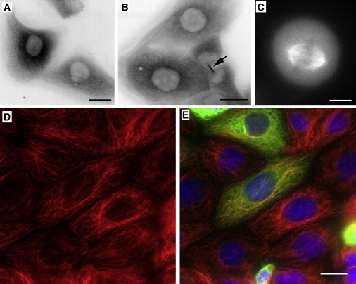
The critical importance of cytoskeletal function for correct neuronal migration during development of the cerebral cortex has been underscored by the identities of germline mutations underlying a number of human neurodevelopmental disorders. The proteins affected include TUBA1A, a major alpha-tubulin isoform, and microtubule-associated components such as doublecortin, and LIS1. Mutations in these genes are associated with the anatomical abnormality lissencephaly, which is believed to reflect failure of neuronal migration. An important recent observation has been the dependence of cortical neuronal migration upon acetylation of alpha-tubulin at lysine 40 by the histone acetyltransferase Elongator complex. Here, we describe a recognizable autosomal recessive syndrome, characterized by generalized polymicrogyria in association with optic nerve hypoplasia (PMGOH). By autozygosity mapping, we show that the molecular basis for this condition is mutation of the TUBA8 gene, encoding a variant alpha-tubulin of unknown function that is not susceptible to the lysine 40 acetylation that regulates microtubule function during cortical neuron migration. Together with the unique expression pattern of TUBA8 within the developing cerebral cortex, these observations suggest a role for this atypical microtubule component in regulating mammalian brain development.
Figures
Comment in
-
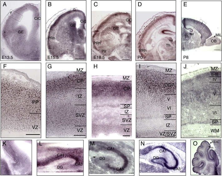
Tuba8 is expressed at low levels in the developing mouse and human brain.Am J Hum Genet. 2010 May 14;86(5):819-22; author reply 822-3. doi: 10.1016/j.ajhg.2010.03.019. Am J Hum Genet. 2010. PMID: 20466094 Free PMC article. No abstract available.
References
-
- Barkovich A.J., Kuzniecky R.I., Jackson G.D., Guerrini R., Dobyns W.B. A developmental and genetic classification for malformations of cortical development. Neurology. 2005;65:1873–1887. - PubMed
-
- Aligianis I.A., Johnson C.A., Gissen P., Chen D., Hampshire D., Hoffmann K., Maina E.N., Morgan N.V., Tee L., Morton J. Mutations of the catalytic subunit of RAB3GAP cause Warburg Micro syndrome. Nat. Genet. 2005;37:221–223. - PubMed
-
- Roll P., Rudolf G., Pereira S., Royer B., Scheffer I.E., Massacrier A., Valenti M.-P., Roeckel-Trevisiol N., Jamali S., Beclin C. SRPX2 mutations in disorders of language cortex and cognition. Hum. Mol. Genet. 2006;15:1195–1207. - PubMed
-
- Glaser T., Ton C.C., Mueller R., Petzl-Erler M.L., Oliver C., Nevin N.C., Housman D.E., Maas R.L. Absence of PAX6 gene mutations in Gillespie syndrome (partial aniridia, cerebellar ataxia, and mental retardation) Genomics. 1994;19:145–148. - PubMed
-
- Baala L., Briault S., Etchevers H.C., Laumonnier F., Natiq A., Amiel J., Boddaert N., Picard C., Sbiti A., Asermouh A. Homozygous silencing of T-box transcription factor EOMES leads to microcephaly with polymicrogyria and corpus callosum agenesis. Nat. Genet. 2007;39:454–456. - PubMed
Publication types
MeSH terms
Substances
Grants and funding
LinkOut - more resources
Full Text Sources
Medical
Molecular Biology Databases
Miscellaneous
