. 2009 Nov;85(5):720-9.
doi: 10.1016/j.ajhg.2009.10.013. Epub 2009 Nov 5.

TRPM1 is mutated in patients with autosomal-recessive complete congenital stationary night blindness

Affiliations

TRPM1 is mutated in patients with autosomal-recessive complete congenital stationary night blindness

Isabelle Audo et al. Am J Hum Genet. 2009 Nov.

Abstract

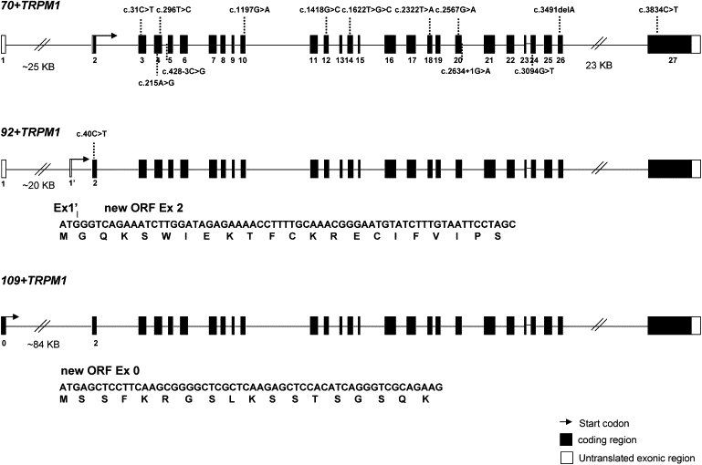
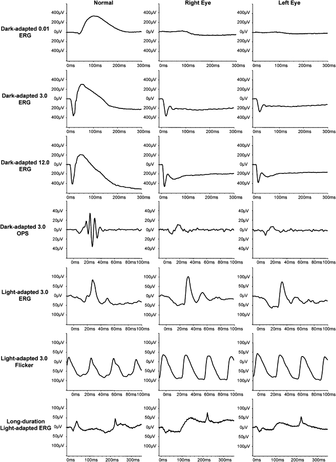
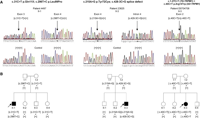
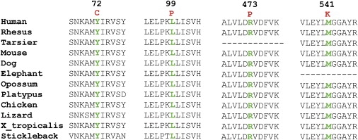
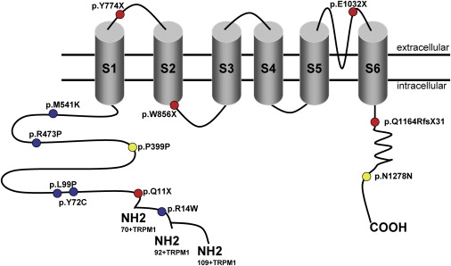
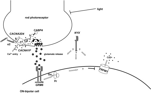
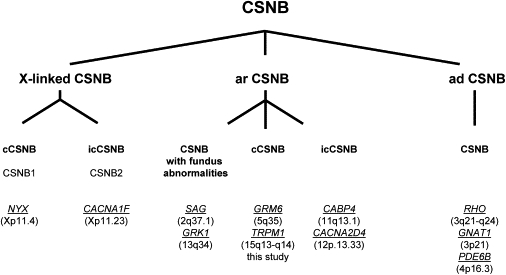
Night vision requires signaling from rod photoreceptors to adjacent bipolar cells in the retina. Mutations in the genes NYX and GRM6, expressed in ON bipolar cells, lead to a disruption of the ON bipolar cell response. This dysfunction is present in patients with complete X-linked and autosomal-recessive congenital stationary night blindness (CSNB) and can be assessed by standard full-field electroretinography (ERG), showing severely reduced rod b-wave amplitude and slightly altered cone responses. Although many cases of complete CSNB (cCSNB) are caused by mutations in NYX and GRM6, in approximately 60% of the patients the gene defect remains unknown. Animal models of human diseases are a good source for candidate genes, and we noted that a cCSNB phenotype present in homozygous Appaloosa horses is associated with downregulation of TRPM1. TRPM1, belonging to the family of transient receptor potential channels, is expressed in ON bipolar cells and therefore qualifies as an excellent candidate. Indeed, mutation analysis of 38 patients with CSNB identified ten unrelated cCSNB patients with 14 different mutations in this gene. The mutation spectrum comprises missense, splice-site, deletion, and nonsense mutations. We propose that the cCSNB phenotype in these patients is due to the absence of functional TRPM1 in retinal ON bipolar cells.


Figures

Figure 1
Figure 1
TRPM1 Isoforms and CSNB Mutations Three isoforms of TRPM1 are presented: the 70+TRPM1 variant represents the previously published reference sequence (RefSeq NM_002420.4), whereas the 92+TRPM1 and 109+TRPM1 isoforms were only recently identified. In comparison to 70+TRPM1, the 92+TRPM1 isoform contains 22 additional amino acids, and the 109+TRPM1 isoform contains 39 additional amino acids. The new open reading frame of 92+TRPM1 is made up of the complete exon 2 with the initiation codon located in a new exon (exon 1′). The new ORF of 109+TRPM1 is made up of the complete exon 2 and a new exon (exon 0). Both new ORFs continue in the isoform 70+TRPM1, coding for 1625 and 1642 amino acids, respectively.
Figure 2
Figure 2
Electrophysiologic Description of Patient CIC00238 with cCSNB, as an Example Full-field ERGs show typical ON bipolar pathway dysfunction: there are no detectable responses for the dark-adapted 0.01 ERG; dark-adapted 3.00 and 12.0 ERGs show an a-wave with a normal amplitude and implicit time but a severely reduced b-wave, leading to an electronegative waveform. Dark-adapted oscillatory potentials (OPs) are not detectable. Light-adapted 3.0 ERGs show normal amplitudes but implicit time shift for both the a-wave and the b-wave. The a-wave has a broadened trough, and there is a sharply rising b-wave with no OPs. Light-adapted 3.0 flicker ERGs show normal amplitudes but a broadened trough and a mildly delayed implicit time. These photopic ERG appearances are characteristic of selective dysfunction of the ON bipolar pathway with OFF bipolar pathway preservation. This is further confirmed with long-duration stimulations, which reveal a normal a-wave but a severely reduced ON-response b-wave and a preserved OFF-response d-wave.
Figure 3
Figure 3
TRPM1 Mutations and Cosegregation Analysis in Families with CSNB (A) Electropherograms of three index patients provided as an example, showing TRPM1 mutations, which are highlighted by an arrow. Exonic sequence is shown in capital letters. Intronic sequence is shown in lowercase letters. (B) Corresponding pedigrees of selected cCSNB patients with TRPM1 mutations and cosegregation in available family members. Filled symbols represent affected individuals, and unfilled symbols represent unaffected persons. Squares indicate males, and circles indicate females. Arrows reflect the index patients.
Figure 4
Figure 4
Evolutionary Conservation of the Altered Amino Acid Residues in Other Orthologs Multiple amino acid sequence alignments show evolutionary conservation of mutated residues (depicted in green). Amino acid substitutions are highlighted in red. The position of the respective amino acids is shown in black numbers.
Figure 5
Figure 5
Localization of TRPM1 Mutations with Respect to Predicted Channel Domains The specific domains for the TRPM1 channel were estimated by the use of different publications and prediction programs (UniProtKB-Swiss-Prot).
Figure 6
Figure 6
Schematic Drawing of Proteins Involved in Signal Transmission from Photoreceptors to Adjacent Bipolar Cells, the Disruption of Which Leads to CSNB Arrows indicate the course of the signal transmission. In darkness, Ca2+ ions enter the rod photoreceptors, which results in glutamate release from the photoreceptors. Activated glutamate receptor activates Gαo1 (arrow), which then closes the TRPM1 channel by an unknown mechanism, indicated by a question mark, and thus ON bipolar cells are hyperpolarized. The exact role of NYX, encoding nyctalopin in this signal transduction cascade, remains to be solved in the future (indicated by a question mark).
Figure 7
Figure 7
Genes Underlying CSNB Different forms of human CSNB are classified according to their mode of inheritance, phenotype, and mutated genes. Abbreviations are as follows: cCSNB, complete CSNB; icCSNB, incomplete CSNB; ar, autosomal recessive; ad, autosomal dominant. Genes are indicated in italics and underlined. Chromosomal location is given between brackets. The phenotype of patients with mutations in icCSNB is more variable and can even lead to progressive cone or cone-rod dystrophy.

References

    1. Zeitz C. Molecular genetics and protein function involved in nocturnal vision. Expert Rev Ophthalmol. 2007;2:467–485.
    1. Schubert G., Bornschein H. Ophthalmologica. 1952;123:396–413. - PubMed
    1. Miyake Y., Yagasaki K., Horiguchi M., Kawase Y., Kanda T. Congenital stationary night blindness with negative electroretinogram. A new classification. Arch. Ophthalmol. 1986;104:1013–1020. - PubMed
    1. Audo I., Robson A.G., Holder G.E., Moore A.T. The negative ERG: clinical phenotypes and disease mechanisms of inner retinal dysfunction. Surv. Ophthalmol. 2008;53:16–40. - PubMed
    1. Zeitz C., Labs S., Lorenz B., Forster U., Ueksti J., Kroes H.Y., De Baere E., Leroy B.P., Cremers F.P., Wittmer M. Genotyping microarray for CSNB-associated genes. Invest Ophthalmol Vis Sci. 2009 Published online July 2, 2009. - PubMed

Publication types