. 2010 Feb 1;242(3):299-309.
doi: 10.1016/j.taap.2009.10.020. Epub 2009 Nov 4.

Nicotine-mediated signals modulate cell death and survival of T lymphocytes

Affiliations

Nicotine-mediated signals modulate cell death and survival of T lymphocytes

Silvia C S Oloris et al. Toxicol Appl Pharmacol. .

Abstract

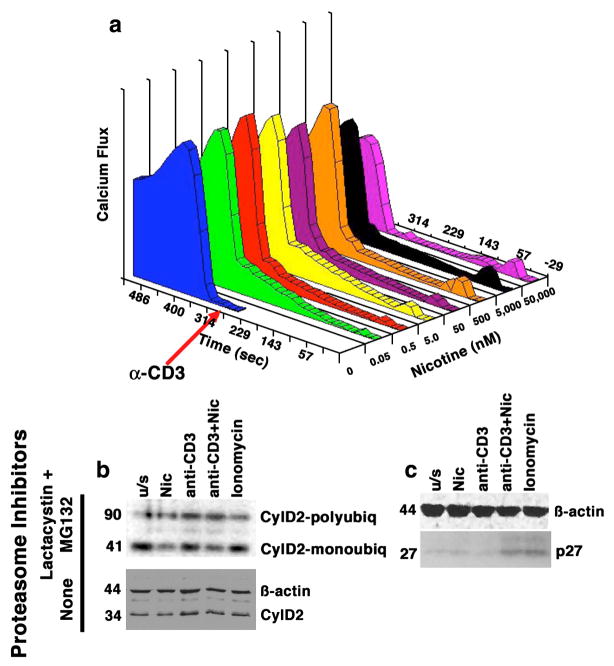
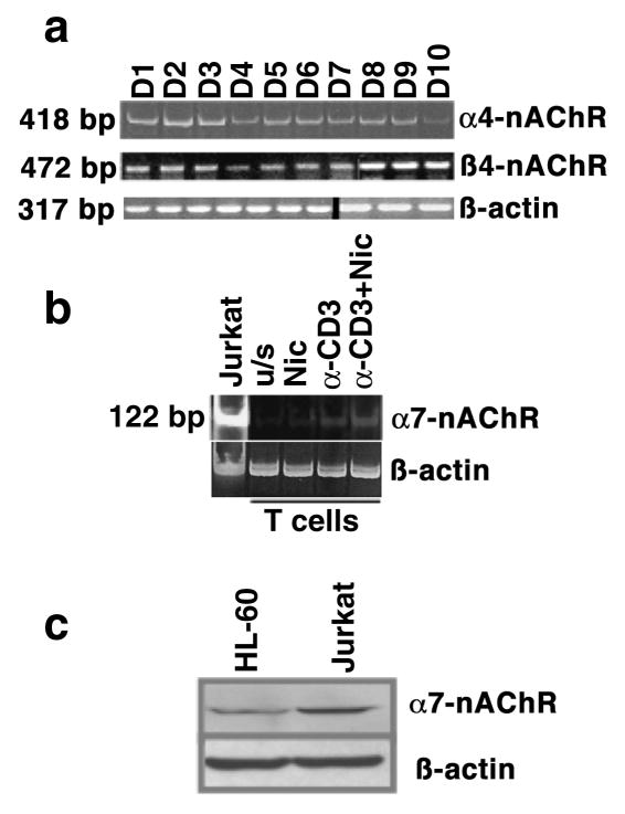
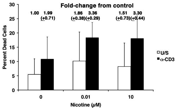
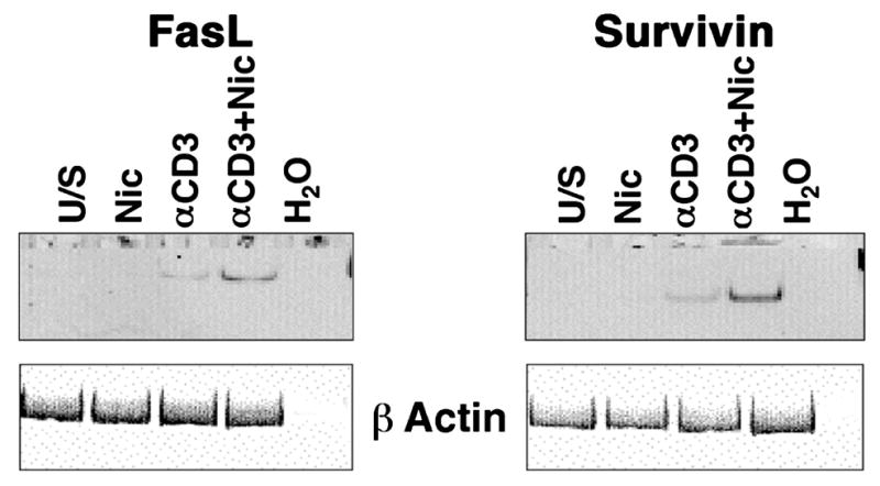
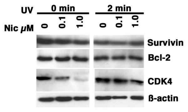
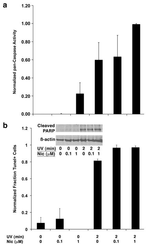
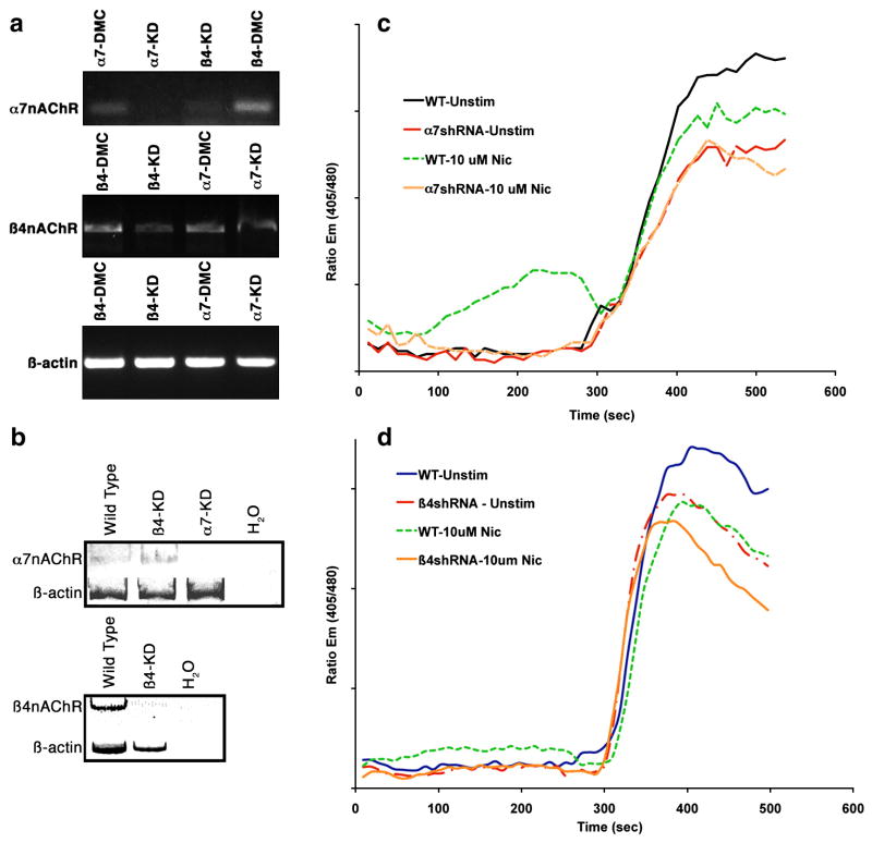
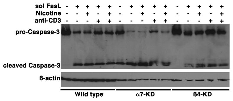
The capacity of nicotine to affect the behavior of non-neuronal cells through neuronal nicotinic acetylcholine receptors (nAChRs) has been the subject of considerable recent attention. Previously, we showed that exposure to nicotine activates the nuclear factor of activated T cells (NFAT) transcription factor in lymphocytes and endothelial cells, leading to alterations in cellular growth and vascular endothelial growth factor production. Here, we extend these studies to document effects of nicotine on lymphocyte survival. The data show that nicotine induces paradoxical effects that might alternatively enforce survival or trigger apoptosis, suggesting that depending on timing and context, nicotine might act both as a survival factor or as an inducer of apoptosis in normal or transformed lymphocytes, and possibly other non-neuronal cells. In addition, our results show that, while having overlapping functions, low and high affinity nAChRs also transmit signals that promote distinct outcomes in lymphocytes. The sum of our data suggests that selective modulation of nAChRs might be useful to regulate lymphocyte activation and survival in health and disease.


Conflict of interest statement

Conflict of Interest: The authors declare that there are no conflicts of interest.

Figures

Figure 1
Figure 1. Dose-dependent calcium mobilization in human T cells induced by nicotine has functional consequences
Panel a shows Jurkat cells treated with nicotine at the indicated concentrations for 5 min prior to the addition of anti-CD3 (10 ng/ml). Alterations in Cai2+ were measured using a MoFlo flow cytometer. Data on the X-axis represent nicotine dose (nM), on the Y-axis, time (sec) and on the Z-axis (Calcium flux) they are expressed as the product of excitation X proportion of responding cells. Panel b shows Jurkat cells cultured as indicated in the presence or absence of proteasome inhibitors Lactacystin and MG132. Cyclin D2 complexes were immunoprecipitated using anti-cyclin D2 antibody and immunoblotted with an anti-ubiquitin antibody (top). Monoubiquitinated Cyclin D2 complexes migrate with an apparent MW of 41 kDa; polyubiquitinated complexes migrate with an apparent MW of ~90 kDa (compared to the 34 kDa native protein). The ratio of polyubiquitinated to monoubiquitinated Cyclin D2 was 2.5-fold and 2-fold greater, respectively, in cells treated with nicotine or with anti-CD3 than in unstimulated cells. The effects of nicotine and anti-CD3 were additive, with the ratio increasing to 3.2-fold over untreated cells when both compounds were used together. The lower immunoblot shows levels of Cyclin D2 in whole cell lysates from cells stimulated in an identical manner without proteasome inhibitors. Panel c shows the levels of the p27 CDK inhibitor in whole cell lysates from primary T cells stimulated in an identical manner in the absence of proteasome inhibitors. The steady state levels of p27 were significantly different (5-fold greater) in cells treated with nicotine and anti-CD3 together, or with ionomycin than in untreated cells. Ionomycin was included in the experiments shown in panel b and panel c to control for non-specific effects of calcium mobilization, and ϐ-actin immunoblots are included as loading controls.
Figure 2
Figure 2. Nicotinic acetylcholine receptor expression in human T lymphocytes
The expression of messenger RNA (a and b) for α4-, ϐ4-, and α7-nAChR subunits, and protein for the α7-subunit (c) was examined in human peripheral blood T cells and in Jurkat T cells using RT-PCR and immunoblotting, respectively. Panel a shows expression of α4- (418 bp amplification product) and ϐ4- (472 bp amplification product) subunits in lymphocytes from ten healthy, adult non-smokers, as well as ϐ-actin as a loading control (note that data for ϐ4-nAChR and for ϐ-actin are compiled from two gels, representing the indicated donors); panel b shows expression of the α7-subunit (122 bp amplification product) in peripheral blood T cells from one representative healthy non-smoker and in Jurkat T cells. ϐ-actin expression in the same samples was used to confirm integrity of the RNA and equivalent loading; panel c shows protein expression of the α7-nAChR in HL-60 and Jurkat cells. A ϐ-actin immunoblot from the same samples is shown as a loading control.
Figure 3
Figure 3. Increased numbers of dead cells are present in activated T cells cultured in the presence of nicotine
Peripheral blood T cells were incubated with or without nicotine for 30 min prior to competence induction with anti-CD3 (10 ng/ml) as indicated and cultured for 48–55 hr without additional stimuli. Loss of cell viability was determined flow cytometrically by uptake of 7-AAD and is indicated as the percent of total events analyzed. The fold-change in dead cells (normalized to 1.00 for unstimulated cells without exposure to nicotine) is shown above each bar. Data represent means (±S.D) from three independent experiments.
Figure 4
Figure 4. Nicotine induces FasL and Survivin gene expression in normal human peripheral blood T cells
Human peripheral blood T cells were incubated with or without nicotine for 30 min prior to competence induction as indicated and cultured for 72 hr without additional stimuli. Expression of FasL and Survivin was examined using RT-PCR under conditions of linear amplification. Amplification products for each gene were 239 and 199 bp, respectively. The same reaction omitting input cDNA (H2O) was used as a negative control. Expression of ϐ-actin (317 bp amplification product) was used to ensure integrity of the RNA. The experiment was repeated in at least 3 donors for each gene with similar results.
Figure 5
Figure 5. Effect of nicotine on expression of Survivin, Bcl-2, and CDK4 in UV-treated Jurkat cells
Jurkat cells were treated by exposure to UV light (2 min) with or without nicotine as indicated and cultured for 4 hours without additional stimuli. Steady state levels of Survivin, Bcl-2, and CDK4 were examined by immunoblotting. ϐ-actin was used as a loading control. The strips shown are from one representative experiment of at least 5 done for each target protein.
Figure 6
Figure 6. Nicotine upregulates caspase activity, but does not tilt the net balance apoptosis in Jurkat cells exposed to UV irradiation
Jurkat cells were treated by exposure to UV light (2 min) with or without nicotine as indicated and cultured for 4 hours without additional stimuli. (A) Pan-caspase activity was analysed fluorimetrically using a FITC-labeled VAD-fmk conjugate. Data show the means (±S.E.M.) of 5 experiments were relative fluorescence was normalized to a maximum of 1.0. (B) DNA fragmentation was measured flow cytometrically using the Apo-BrDU Tunel assay kit. Data show the means (±S.E.M.) of 3 experiments were the maximum rate of apoptosis (97% for UV + nicotine-treated cells) was normalized to 1.0. The inset shows immunoblots from whole cell lysates were evaluating cleavage of PARP (a prototypical Caspase-3 substrate). ϐ-actin was used as a loading control.
Figure 7
Figure 7. Attenuation of nAChRs in Jurkat cells using RNA interference
Panel a shows the effect of transient transfection of shRNA constructs for ϐ4- (Beta-4.5) or α7- (Alpha-7.13) nAChR subunits in Jurkat T cells. Controls were designed for each shRNA by creating double point mutant oligonucleotides (see Table 2). Amplification products for each subunit and for ϐ-actin (as a control for RNA integrity and loading) shown in the figure are from the same experiment. Control shRNA vectors are indicated by the abbreviation DMC (double point mutant control). Panel b illustrates the nAChR expression phenotypes in Jurkat T cells after puromycin selection. Expression of nAChR mRNA in both transiently and stably transfected cells was examined by RT-PCR under conditions of linear amplification (KD = “knockdown”). Panels c and d show calcium mobilization in the nAChR-knockdown Jurkat cells. Control Jurkat cells (“WT”), α7-KD (“a7shRNA”), or ϐ4-KD cells (“ϐ4shRNA) cells were left untreated or treated with nicotine (10 μM) as indicated. After 5 min, cells were allowed to remain as previously treated, or were stimulated by addition of anti-CD3. Alterations in intracellular ionized calcium (Cai++) were measured using a MoFlo flow cytometer. Y-axis values represent Indo-1 emission ratios at 405 nm/480 nm. Basal ionized calcium levels were ~150 nM.
Figure 8
Figure 8. Effect of nAChR loss on Jurkat cell apoptosis
Control Jurkat cells (WT),α7-nAChR knockdowns (α7-KD) or ϐ4-nAChR-knockdowns (ϐ4-KD) were cultured in the presence or absence of soluble FasL (sFasL) for 4 hr as indicated, with or without a 15-min pre-exposure to nicotine (1 μM) and/or concomitant exposure to anti-CD3 (10 ng/ml). Cleavage of pro-Caspase-3, examined by immunoblotting, was used as a measure of apoptosis. ϐ-actin was used as a loading control. The data show one representative experiment of three done.
Figure 9
Figure 9. Effect of nAChR loss on survival of normal human peripheral blood T cells
T cells were transfected with dsRed and the shRNA constructs indicated. Transfection efficiencies were comparable for all conditions after 24 hr. The number of dsRed+ cells remaining in the cultures was evaluated by flow cytometry after 72 hr. Mean percent (range) dsRed+ cells in triplicate transfections were: none (background), 2.6% (2.4–2.8); Control, 31.7%(25–34), ϐ4-nAChR shRNA, 28%(26–30); α7-nAChR shRNA, 17.4%(15–19); ϐ4-nAChR shRNA+α7 nAChR shRNA, 1.8%(1.3–2.5).

References

    1. Abraham RT, Weiss A. Jurkat T cells and development of the T-cell receptor signalling paradigm. Nat Rev Immunol. 2004;4:301–308. - PubMed
    1. Amos CI, Wu X, Broderick P, Gorlov IP, Gu J, Eisen T, Dong Q, Zhang Q, Gu X, Vijayakrishnan J, Sullivan K, Matakidou A, Wang Y, Mills G, Doheny K, Tsai YY, Chen WV, Shete S, Spitz MR, Houlston RS. Genome-wide association scan of tag SNPs identifies a susceptibility locus for lung cancer at 15q25.1. Nat Genet. 2008;40:616–622. - PMC - PubMed
    1. Aoshiba K, Nagai A, Yasui S, Konno K. Nicotine prolongs neutrophil survival by suppressing apoptosis. J Lab Clin Med. 1996;127:186–194. - PubMed
    1. Arredondo J, Hall LL, Ndoye A, Nguyen VT, Chernyavsky AI, Bercovich D, Orr-Urtreger A, Beaudet AL, Grando SA. Central role of fibroblast alpha3 nicotinic acetylcholine receptor in mediating cutaneous effects of nicotine. Lab Invest. 2003;83:207–225. - PubMed
    1. Arredondo J, Nguyen VT, Chernyavsky AI, Bercovich D, Orr-Urtreger A, Kummer W, Lips K, Vetter DE, Grando SA. Central role of alpha7 nicotinic receptor in differentiation of the stratified squamous epithelium. J Cell Biol. 2002;159:325–336. - PMC - PubMed

Publication types

MeSH terms