. 2010 Jan;152(1):151-65.
doi: 10.1104/pp.109.145656. Epub 2009 Nov 6.

The Arabidopsis tandem zinc finger protein AtTZF1 traffics between the nucleus and cytoplasmic foci and binds both DNA and RNA

Affiliations

The Arabidopsis tandem zinc finger protein AtTZF1 traffics between the nucleus and cytoplasmic foci and binds both DNA and RNA

Marcelo C Pomeranz et al. Plant Physiol. 2010 Jan.

Abstract

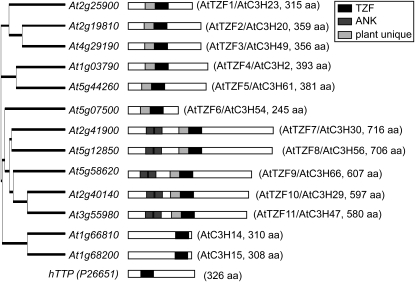
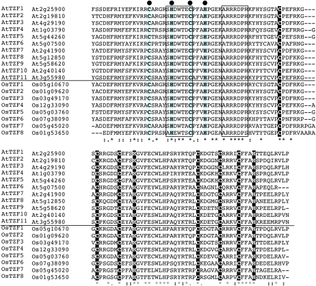
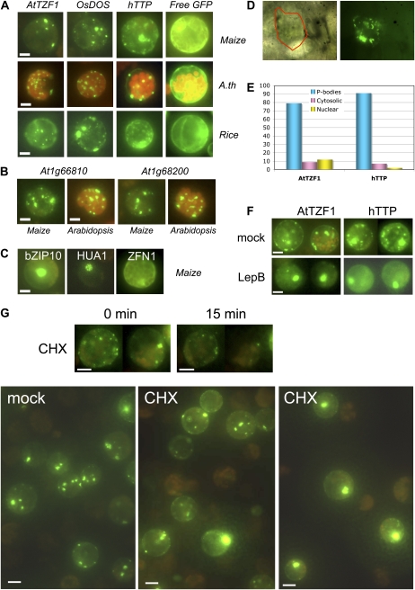
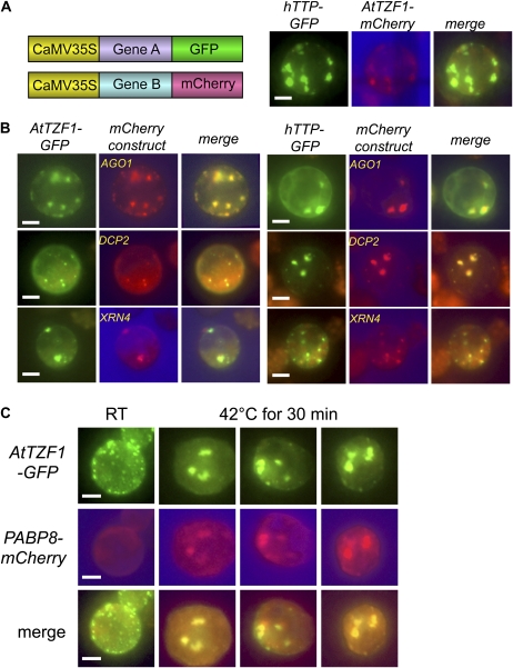
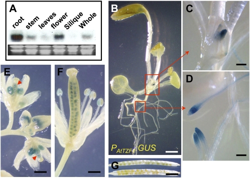
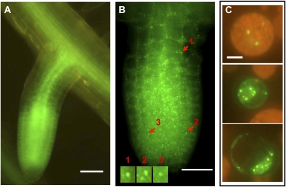
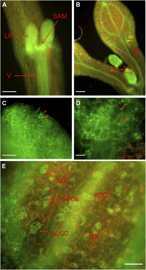
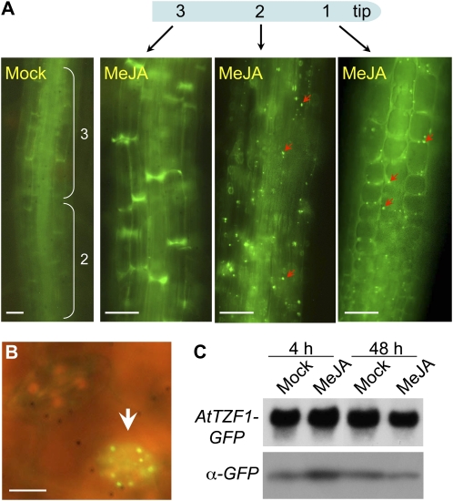
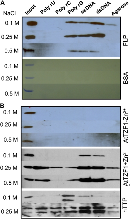
Processing bodies (PBs) are specialized cytoplasmic foci where mRNA turnover and translational repression can take place. Stress granules are related cytoplasmic foci. The CCCH tandem zinc finger proteins (TZFs) play pivotal roles in gene expression, cell fate specification, and various developmental processes. Human TZF binds AU-rich elements at the 3' untranslated region and recruits decapping, deadenylation, and exonucleolytic enzymes to PBs for RNA turnover. Recent genetic studies indicate that plant TZFs are involved in gene regulation and hormone-mediated environmental responses. It is unknown if plant TZFs can bind RNA and be localized to PBs or stress granules. The Arabidopsis (Arabidopsis thaliana) AtTZF1/AtCTH/AtC3H23 was identified as a sugar-sensitive gene in a previous microarray study. It is characterized by a TZF motif that is distinct from the human TZF. Higher plants such as Arabidopsis and rice (Oryza sativa) each have a gene family containing this unique TZF motif. Here, we show that AtTZF1 can traffic between the nucleus and cytoplasmic foci. AtTZF1 colocalizes with markers of PBs, and the morphology of these cytoplasmic foci resembles that of mammalian PBs and stress granules. AtTZF1-associated cytoplasmic foci are dynamic and tissue specific. They can be induced by dark and wound stresses and are preferentially present in actively growing tissues and stomatal precursor cells. Since AtTZF1 can bind both DNA and RNA in vitro, it raises the possibility that AtTZF1 might be involved in DNA and/or RNA regulation.


Figures

Figure 1.
Figure 1.
Phylogenetic analysis of the AtTZF gene family. All AtTZF genes are characterized by the C-x7-C-x5-C-x3-H-x16-C-x5-C-x4-C-x3-H motif, except that At1g03790 and At5g44260 contain the variant C-x8-C-x5-C-x3-H-x16-C-x5-C-x4-C-x3-H motif. Also shown are At1g66810 and At1g68200, which contain the TZF motif C-x8-C-x5-C-x3-H-x18-C-x8-C-x5-C-x3-H, identical to the TZF in hTTP. Nomenclatures with the AtC3H symbols (Wang et al., 2008) are included for cross-reference. ANK, Ankyrin repeat.
Figure 2.
Figure 2.
Plant-unique TZF motif. Sequence comparison of the highly conserved CCCH TZF motif (white letters on black background) and upstream 50 amino acids of Arabidopsis AtTZF and rice OsTZF genes using the ClustalW program (http://www.ebi.ac.uk/Tools/clustalw2/index.html). A conserved C-x5-H-x4-C-x3-H motif is highlighted by black dots, and two other conserved motifs, SHDWTEC and ARRRDPR, are boxed. [See online article for color version of this figure.]
Figure 3.
Figure 3.
AtTZF1 is localized in the cytoplasmic foci that resemble PBs. A, AtTZF1-GFP, rice homolog OsDOS (Os01g09620)-GFP, and hTTP (P26651)-GFP are localized in cytoplasmic foci in etiolated maize, green Arabidopsis (A.th), and etiolated rice leaf mesophyll protoplasts. By contrast, free GFP is localized in the cytoplasm in a diffusive pattern. Bar = 10 μm. B, The closest Arabidopsis homologs of hTTP, At1g66810 and At1g68200, are also predominantly localized in cytoplasmic foci. Bar = 10 μm. C, For comparison, transcription factor bZIP10 (At4g02640)-GFP is localized in the nucleus, HUA1-GFP (At3g12680; six CCCH zinc fingers) is localized in the nucleolus, and ZFN1-GFP (At3g02830; five CCCH zinc fingers) is localized in the cytoplasm. Bar = 10 μm. D, AtTZF1-GFP is localized in the cytoplasmic foci in lima bean cotyledon cells transformed via particle bombardment. A single cell is outlined in red in a corresponding transmitting bright-light image (left). E, Differential subcellular localization of AtTZF1 and hTTP in maize protoplasts. Shown are the percentages of cells (AtTZF1, 669 cells; hTTP, 376 cells) with respect to the pattern indicated. F, AtTZF1 and hTTP can shuttle between cytoplasmic foci and the nucleus. Shown are maize protoplasts that express either AtTZF1-GFP or hTTP-GFP in the absence/presence of leptomycin B (LepB), an inhibitor of nuclear export receptor. Bar = 10 μm. G, Negative effects of CHX on cytoplasmic foci formation. The number and intensity of cytoplasmic foci are reduced by the CHX treatment in 15 min (top panel). However, some cytoplasmic foci are relatively insensitive to CHX (bottom middle panel), indicating their non-PB/SG identities. By contrast, some CHX-treated cells show GFP signals in the nucleus (bottom right panel), indicating that CHX is effective in reducing AtTZF1-associated cytoplasmic foci but is ineffective when AtTZF1 is localized in the nucleus. Bar = 10 μm.
Figure 4.
Figure 4.
AtTZF1 is colocalized with PB and SG markers. A, Constructs used for the colocalization studies, and colocalization results of hTTP and AtTZF1 in maize protoplasts. Bar = 10 μm. B, Both AtTZF1 and hTTP are colocalized with PB markers AGO1, DCP2, and XRN4. Bar = 10 μm. C, AtTZF1 can be localized in the SGs. Both AtTZF1 and putative SG marker PABP8 form aggregates and colocalize after heat shock at 42°C for 30 min. Bar = 10 μm. RT, Room temperature.
Figure 5.
Figure 5.
Temporal and spatial expression of AtTZF1. A, RNA gel-blot analysis indicates that AtTZF1 mRNA is differentially expressed in different tissues. B, GUS staining showing differential expression of PAtTZF1:GUS in root tips and vasculature. Bar = 2 mm. C, Enlarged view of shoot apical region showing GUS stain in young leaf primordia. Bar = 200 μm. D, Enlarged view showing GUS stain in root tips and vasculature. Bar = 200 μm. E, PAtTZF1:GUS is expressed in anthers and female gametophytes (arrowheads) before pollination. Bar = 0.5 mm. F and G, Differential expression of PAtTZF1:GUS during embryo development. Shown are high levels of PAtTZF1:GUS expression in early stages of embryo development (F; bar = 0.5 mm) and very low to no expression after bending-cotyledon stages (G; bar = 2.5 mm).
Figure 6.
Figure 6.
Subcellular localization of AtTZF1 in CaMV35S:AtTZF1-GFP plants. A, AtTZF1-associated cytoplasmic foci are most commonly found in the root meristems, where the protein is accumulated at higher levels. B, Closeup view of a root tip showing cytoplasmic foci within each cell. Bar = 50 μm. Boxed regions are shown with enlarged views. C, Cytoplasmic foci can be found in the leaf protoplasts isolated from CaMV35S:AtTZF1-GFP plants. Bar = 10 μm.
Figure 7.
Figure 7.
AtTZF1-associated cytoplasmic foci are present in etiolated seedlings and can be induced by wounding. A, AtTZF1-GFP is highly abundant in leaf primordia (LP), shoot apical meristem (SAM), and vasculature (V). B, In contrast to the fully expanded cotyledons, where AtTZF1-GFP is localized mainly in the vasculature, it is present in all cells in the true leaf primordia. In addition, AtTZF1-GFP cytoplasmic foci can be induced in the cells surrounding a wound (W) pinched by a pair of fine-tip forceps. Bar = 100 μm. C, Enlarged view is shown from the red rectangular area in B, a leaf primordium with numerous cytoplasmic foci. Some cytoplasmic foci are indicated by arrows. Bar = 20 μm. D, Enlarged view is shown from the red rectangular area in B, showing an induction of cytoplasmic foci in the cells surrounding a wounding site (bottom right). Some cytoplasmic foci are indicated by arrows. Bar = 10 μm. E, In etiolated cotyledons, AtTZF1-associated cytoplasmic foci are especially distinct in stomatal precursor cells (SPCs), in young guard cells (YGCs), and in stomatal-lineage ground cells (SLGCs) but are not apparent in mature stomata (MS). Bar = 30 μm.
Figure 8.
Figure 8.
Wound hormone MeJA induces cytoplasmic foci formation. A, Cytoplasmic foci are normally found in root meristems. However, they can be found in transition (1) and elongation-differentiation (2) zones (arrowheads) after the MeJA (100 μm) treatment. Also shown are mock-treated root zone 2 and fully elongated zone 3. Bar = 50 μm. B, Cytoplasmic foci can be induced by MeJA in most stomata (arrowhead). Bar = 10 μm. C, Induction of cytoplasmic foci is unlikely a direct consequence of increased protein levels. RNA gel blot shows similar expression levels between mock- and MeJA-treated plants. The protein level is higher only in 4-h MeJA-treated samples. Images in A and B were taken from 48-h samples. [See online article for color version of this figure.]
Figure 9.
Figure 9.
AtTZF1 binds both DNA and RNA in vitro. A, In vitro bead-binding assays showing that the MYB transcription factor FLP preferentially binds DNA. Binding to the poly-rG appears to be nonspecific. As a negative control, BSA binds neither DNA nor RNA. B, Results of in vitro bead-binding assays indicate that AtTZF1 preferentially binds single- and double-stranded DNA at 0.1 m NaCl and can bind ribohomopolymer U when salt concentration increases to 0.25 to 0.5 m. Poly-rG and poly-rC binding appears to be nonspecific, as the negative control (agarose) can generate similar signals. The binding requires the presence of Zn2+, as no binding is detected in the absence of ZnCl2 in the binding buffer. In contrast to AtTZF1, hTTP not only binds both single-stranded and double-stranded DNA but also binds all ribohomopolymers with similar affinities at 0.5 m NaCl. The results by poly-rG are likely false positive, because every protein tested so far can generate similar signals (data not shown). [See online article for color version of this figure.]
Figure 10.
Figure 10.
AtTZF1 does not bind the hTTP consensus binding site. A, REMSA shows that hTTP can bind the 3′ UTR of TNF-α with specificity, because mutation in either RNA probe (MutG) or TZF domain (C124R) can eliminate the binding (indicated by the arrow). By contrast, no RNA-protein complex can be detected when AtTZF1 is used. B, REMSA results indicate that neither hTTP nor AtTZF1 can bind the GASA6 3′ UTR. The arrowhead indicates free probe.

Comment in

  • AtTZF gene family localizes to cytoplasmic foci.
    Pomeranz M, Lin PC, Finer J, Jang JC. Pomeranz M, et al. Plant Signal Behav. 2010 Feb;5(2):190-2. doi: 10.4161/psb.5.2.10988. Epub 2010 Feb 18. Plant Signal Behav. 2010. PMID: 20173417 Free PMC article.

Similar articles

Cited by

References

    1. Anderson P, Kedersha N (2006) RNA granules. J Cell Biol 172 803–808 - PMC - PubMed
    1. Anderson P, Kedersha N (2008) Stress granules: the tao of RNA triage. Trends Biochem Sci 33 141–150 - PubMed
    1. Balagopal V, Parker R (2009) Polysomes, P bodies and stress granules: states and fates of eukaryotic mRNAs. Curr Opin Cell Biol 21 403–408 - PMC - PubMed
    1. Barbee SA, Estes PS, Cziko AM, Hillebrand J, Luedeman RA, Coller JM, Johnson N, Howlett IC, Geng C, Ueda R, et al (2006) Staufen- and FMRP-containing neuronal RNPs are structurally and functionally related to somatic P bodies. Neuron 52 997–1009 - PMC - PubMed
    1. Barreau C, Dutertre S, Paillard L, Osborne HB (2006) Liposome-mediated RNA transfection should be used with caution. RNA 12 1790–1793 - PMC - PubMed

Publication types

MeSH terms

LinkOut - more resources