. 2010 Jan;192(2):502-17.
doi: 10.1128/JB.00803-09. Epub 2009 Nov 6.

A genomic island defines subspecies-specific virulence features of the host-adapted pathogen Campylobacter fetus subsp. venerealis

Affiliations

A genomic island defines subspecies-specific virulence features of the host-adapted pathogen Campylobacter fetus subsp. venerealis

Gregor Gorkiewicz et al. J Bacteriol. 2010 Jan.

Abstract

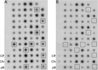
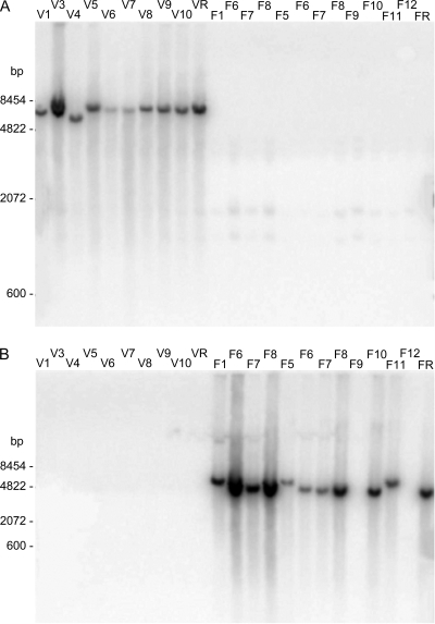
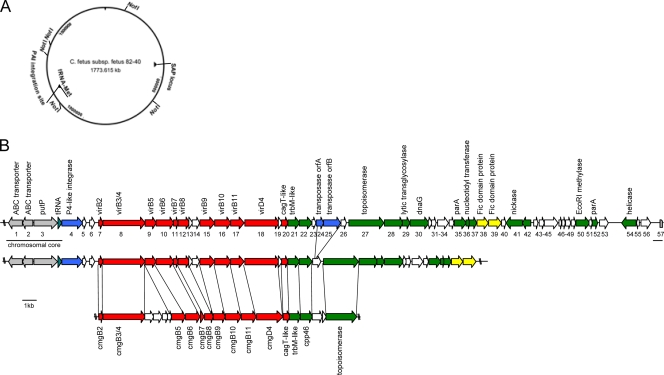
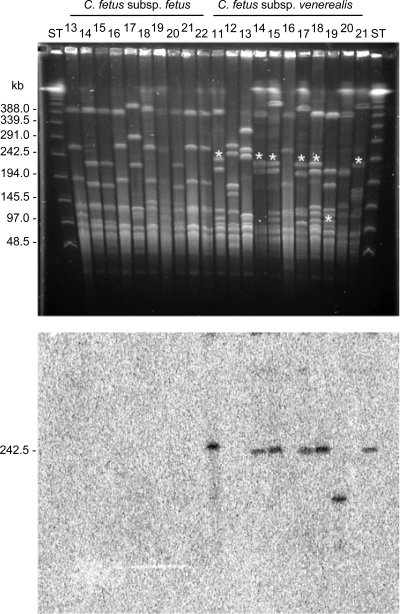
The pathogen Campylobacter fetus comprises two subspecies, C. fetus subsp. fetus and C. fetus subsp. venerealis. Although these taxa are highly related on the genome level, they are adapted to distinct hosts and tissues. C. fetus subsp. fetus infects a diversity of hosts, including humans, and colonizes the gastrointestinal tract. In contrast, C. fetus subsp. venerealis is largely restricted to the bovine genital tract, causing epidemic abortion in these animals. In light of their close genetic relatedness, the specific niche preferences make the C. fetus subspecies an ideal model system to investigate the molecular basis of host adaptation. In this study, a subtractive-hybridization approach was applied to the genomes of the subspecies to identify different genes potentially underlying this specificity. The comparison revealed a genomic island uniquely present in C. fetus subsp. venerealis that harbors several genes indicative of horizontal transfer and that encodes the core components necessary for bacterial type IV secretion. Macromolecular transporters of this type deliver effector molecules to host cells, thereby contributing to virulence in various pathogens. Mutational inactivation of the putative secretion system confirmed its involvement in the pathogenicity of C. fetus subsp. venerealis.


Figures

FIG. 1.
FIG. 1.
RDA with C. fetus subsp. venerealis DNA as tester DNA reveals candidate clones for subspecies-specific genomic fragments. Typical results for the analysis are illustrated with duplicate dot blots of 71 clones. Blot A was hybridized with Sau3A1-digested and 32P-labeled genomic C. fetus subsp. venerealis ATCC 19438 DNA. Blot B was hybridized with an equivalently prepared C. fetus subsp. fetus ATCC 27374 genomic DNA probe. The squares indicate clones specific for the C. fetus subsp. venerealis reference strain (no hybridization with the C. fetus subsp. fetus probe). DNA (500 ng) from reference strains were applied as positive controls (Cff and Cfv) as marked, and vector DNA (300 ng) was used to control for plasmid hybridization (pB).
FIG. 2.
FIG. 2.
Distribution of hybridization signals indicating the subspecies specificity of genomic fragments 3Dv and 4Ch. Genomic DNA (4 μg) from a selection of reference and field strains of C. fetus was digested with 20 U HaeIII, resolved on 1% agarose Tris-acetate-EDTA (TAE) gels, and transferred to nitrocellulose for Southern analysis. The identification numbers above the lanes designate isolates of the strain collection, as listed in Table 1. Samples F6 to F8 were loaded twice. The first set (left) contained 5-fold more genomic DNA. (A) A 32P-labeled probe derived from clone 3Dv hybridized specifically to complementary sequences exclusively present in C. fetus subsp. venerealis strains (lanes V1 to VR). (B) A 32P-labeled-probe derived from clone 4Ch hybridized to genomic DNA from the majority of C. fetus subsp. fetus strains tested (lanes F1 to FR), but not to DNA from C. fetus subsp. venerealis strains (lanes V1 to VR).
FIG. 3.
FIG. 3.
Schematic representation of the C. fetus subsp. venerealis-specific genomic island. (A) The site of integration of the genomic island (PAI) is projected onto a map of the sequenced genome of C. fetus subsp. fetus 82-40 in the vicinity of a methionyl-tRNA gene. The SAP locus denotes the localization of the surface layer protein locus. (B) Annotated genomic island of C. fetus subsp. venerealis ATCC 19438 (top) and data from the partially completed C. fetus subsp. venerealis 84-112 (V81) genome (middle) compared to the highly syntenic resistance plasmid pCC31 of C. coli (bottom). The site of island insertion into the chromosomal core (gray, underlined) follows a tRNA gene. Gene designations are given as numbers under the gene map, and putative functional assignments are shown above. The corresponding functional groups are highlighted with color, including mobility genes, such as IS transposases and phage integrases (blue), a type IV secretion system (red), putative effector molecules (yellow), genes of apparent plasmid origin (green), and genes of unknown function (white). The scale bar represents 1 kb.
FIG. 4.
FIG. 4.
The C. fetus subsp. venerealis genomic island exhibits a conserved position of chromosome insertion. PFGE of SmaI-digested C. fetus DNA (top), followed by Southern hybridization with a radioactively labeled orf21 probe (bottom), reveals a typical resolution of the unique gene region on a 240-kb fragment. The asterisks (top) identify hybridized fragments. The identification numbers above the lanes designate isolates of the strain collection, as listed parenthetically in Table S1 in the supplemental material. A PFGE-DNA ladder (New England Biolabs) served as a standard (ST).
FIG. 5.
FIG. 5.
Comparative analysis of C. fetus subsp. venerealis ATCC 19438 (CfvT) and the virB9 mutant (Cfv 3-18) vir gene expression. Shown are reverse transcriptase-PCR products generated from cDNA obtained from the strains indicated (right) for the targeted genes shown (top). Control reactions included genomic DNA (DNA) and DNase-treated RNA without reverse transcription (RNA), as indicated above the lanes. Products (10 μl) were separated on a 2% agarose-TAE gel with 500 ng of λ HindIII marker and 1 μg of a 100-bp DNA ladder (st). Primers (Table 2) hybridized as follows: (1) VirB8-4 and VirB8-14 within virB8; (2) UP4 and VirB9-2 in virB9 or (3) VirB9_HindIII_rev and VirB9_PstI_fwd within virB9 and flanking the insertion site of aphA-3; (4) KmR_rev and VirB9_KmR_fwd, spanning from virB9 to aphA-3; (5) UP3 and UP1 in virB10 and (6) TaxB3 and TaxB2 in virD4.
FIG. 6.
FIG. 6.
The virB9 mutant Cfv 3-18 shows an impaired cytotoxic phenotype in continuous coculture with HeLa cells. HeLa cells were grown to confluence and then infected in triplicate at an MOI of 100 with either C. fetus subsp. venerealis ATCC 19438 (CfvT) or Cfv 3-18. (A) The number of surviving HeLa cells relative to uninfected control cultures is indicated. (B) The number of viable bacteria in the cell culture medium was determined daily. No statistical difference between CfvT and Cfv 3-18 was observed. The values shown are averages of 3 independent experiments. Statistical significance is indicated: *, P < 0.05; **, P < 0.005 (CfvT versus Cfv 3-18). The error bars indicate standard deviations.
FIG. 7.
FIG. 7.
virD4 mutation attenuates bacterial invasion. (A) Caco-2 cells were infected with C. fetus subsp. venerealis 84-112 harboring the vector control (left) (V81[pRYSS1]), the isogenic virD4 mutant harboring the vector control (middle) (V81_SK1[pRYSS1]), or the mutant strain harboring virD4 in trans (right) (V81_SK1[pRYVL2]). Extracellular bacteria were killed with gentamicin after 5 h, and the relative number of intracellular bacteria per Caco-2 cell was compared to that of the wild type (WT) (set to 100%). (B) For a negative control of the invasion phenotype, Caco-2 cells were infected with wild-type C. fetus subsp. venerealis 84-112 (V81) or the isogenic mutant in cdtB (V81_JL1) for 3 h. The values shown are the means of three independent experiments plus standard deviations; *, P < 0.02.

Similar articles

Cited by

References

    1. Amieva, M. R., R. Vogelmann, A. Covacci, L. S. Tompkins, W. J. Nelson, and S. Falkow. 2003. Disruption of the epithelial apical-junctional complex by Helicobacter pylori CagA. Science 300:1430-1434. - PMC - PubMed
    1. Anonymous. 1996. OIE manual of standards for diagnostic tests and vaccines, 3rd ed., p. 256-266. Office International des Epizooties, Paris, France.
    1. Asakura, M., W. Samosornsuk, M. Taguchi, K. Kobayashi, N. Misawa, M. Kusumoto, K. Nishimura, A. Matsuhisa, and S. Yamasaki. 2007. Comparative analysis of cytolethal distending toxin (cdt) genes among Campylobacter jejuni, C. coli and C. fetus strains. Microb. Pathog. 42:174-183. - PubMed
    1. Ausubel, F. M., R. Brent, R. E. Kingston, D. D. Moore, J. A. Smith, J. G. Seidman, and K. Struhl. 1987. Current protocols in molecular biology. John Wiley & Sons, New York, NY.
    1. Baar, C., M. Eppinger, G. Raddatz, J. Simon, C. Lanz, O. Klimmek, R. Nandakumar, R. Gross, A. Rosinus, H. Keller, P. Jagtap, B. Linke, F. Meyer, H. Lederer, and S. C. Schuster. 2003. Complete genome sequence and analysis of Wolinella succinogenes. Proc. Natl. Acad. Sci. U. S. A. 100:11690-11695. - PMC - PubMed

Publication types

MeSH terms

Substances

Associated data

LinkOut - more resources