. 2009 Oct 29;4(10):e7630.
doi: 10.1371/journal.pone.0007630.

Chromosome 7 and 19 trisomy in cultured human neural progenitor cells

Affiliations

Chromosome 7 and 19 trisomy in cultured human neural progenitor cells

Dhruv Sareen et al. PLoS One. .

Abstract

Background: Stem cell expansion and differentiation is the foundation of emerging cell therapy technologies. The potential applications of human neural progenitor cells (hNPCs) are wide ranging, but a normal cytogenetic profile is important to avoid the risk of tumor formation in clinical trials. FDA approved clinical trials are being planned and conducted for hNPC transplantation into the brain or spinal cord for various neurodegenerative disorders. Although human embryonic stem cells (hESCs) are known to show recurrent chromosomal abnormalities involving 12 and 17, no studies have revealed chromosomal abnormalities in cultured hNPCs. Therefore, we investigated frequently occurring chromosomal abnormalities in 21 independent fetal-derived hNPC lines and the possible mechanisms triggering such aberrations.

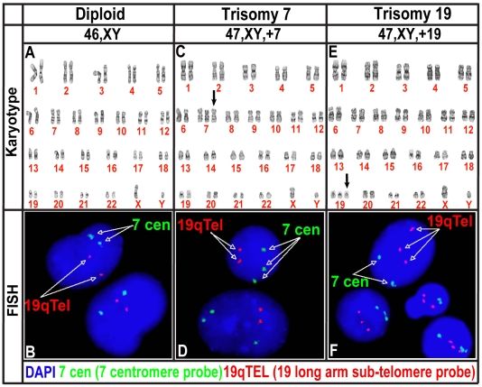
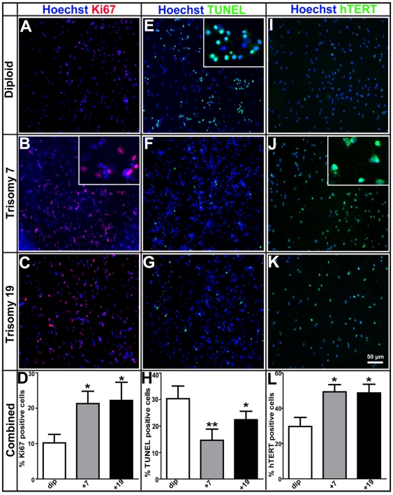
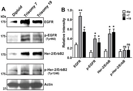
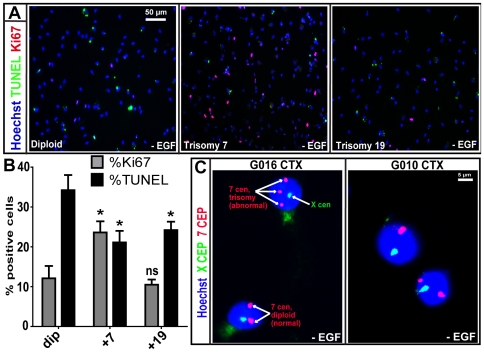
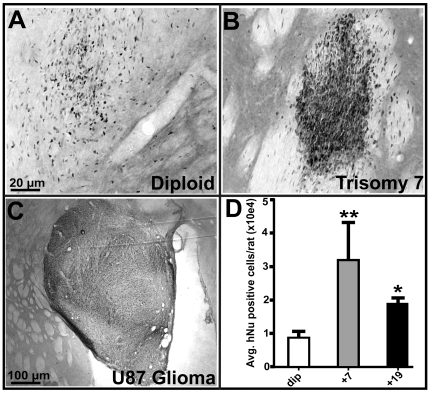
Methods and findings: While most hNPC lines were karyotypically normal, G-band karyotyping and fluorescent in situ hybridization (FISH) analyses revealed the emergence of trisomy 7 (hNPC(+7)) and trisomy 19 (hNPC(+19)), in 24% and 5% of the lines, respectively. Once detected, subsequent passaging revealed emerging dominance of trisomy hNPCs. DNA microarray and immunoblotting analyses demonstrate epidermal growth factor receptor (EGFR) overexpression in hNPC(+7) and hNPC(+19) cells. We observed greater levels of telomerase (hTERT), increased proliferation (Ki67), survival (TUNEL), and neurogenesis (beta(III)-tubulin) in hNPC(+7) and hNPC(+19), using respective immunocytochemical markers. However, the trisomy lines underwent replicative senescence after 50-60 population doublings and never showed neoplastic changes. Although hNPC(+7) and hNPC(+19) survived better after xenotransplantation into the rat striatum, they did not form malignant tumors. Finally, EGF deprivation triggered a selection of trisomy 7 cells in a diploid hNPC line.

Conclusions: We report that hNPCs are susceptible to accumulation of chromosome 7 and 19 trisomy in long-term cell culture. These results suggest that micro-environmental cues are powerful factors in the selection of specific hNPC aneuploidies, with trisomy of chromosome 7 being the most common. Given that a number of stem cell based clinical trials are being conducted or planned in USA and a recent report in PLoS Medicine showing the dangers of grafting an inordinate number of cells, these data substantiate the need for careful cytogenetic evaluation of hNPCs (fetal or hESC-derived) before their use in clinical or basic science applications.


Conflict of interest statement

Competing Interests: Dr. Clive N. Svendsen has the following competing interest disclosure for his work with Brain Cells, Inc. a) acted as a consultant within the last 2 years b) performed contract work within the last 2 years c) received more than $10,000 a year from the company within the last 2 years. There are no patents or any other benefits to Brain Cells Inc upon publication of this paper.

Figures

Figure 1
Figure 1. Trisomy of chromosome 7 and 19 in M031 CTX hNPC line.
(A, B) Compared to the wild type controls (dip), (C, D) sub-cultures of M031 CTX line display a complete trisomy of chromosome 7 (+7), (E, F) and a trisomy of chromsome19 (+19) after fourteen to thirty-eight passages. (A, C, E) Twenty metaphase cells were examined by G-banding, (B, D, F) and 200 interphase nuclei were evaluated by fluorescence in-situ hybridization (FISH). Results are representative of at least one of three independent biological samples with similar results.
Figure 2
Figure 2. Growth and proliferative advantage of M031+7 and M031+19 over M031dip CTX hNPCs.
(A–D) Immunocytochemistry for Ki67 protein, a cell-cycle associated proliferation marker, shows significantly more Ki67 positive cells in both M031+7(21%) and M031+19 (22%) compared to the M031dip line (10%). (E–H) Cell death analysis using TUNEL staining shows that compared to the M031dip control (31%), there was a significant decline in the number TUNEL positive cells in the M031+7 (13%) and M031+19 (22%) hNPCs. (I–L) Immunocytochemistry for the catalytic subunit of telomerase (hTERT) exhibits significant increase in the number of hTERT positive cells in M031+7 (49%) and M031+19 (48%), when compared to M031dip controls (∼31%). p value: ** <0.01 and * <0.05. The magnified images for (B) Ki67, (E) TUNEL, and (J) hTERT immunostaining are shown in insets. Images are representative of one of three independent experiments with similar results. The data in the graphs are averaged over three independent experiments with mean and SEM values.
Figure 3
Figure 3. EGF signaling is important in survival advantage of trisomy hNPCs.
(A) Western blot analysis illustrated that M031+7 and M031+19 lines overexpress EGFR family proteins resulting in increased activation of phosphorylated EGFR (Tyr992). (B) Desnitometry quantification of scanned bands using ImageJ 1.17 software displays ∼2 to 3-fold increase in EGFR protein levels and 3 to 4-fold increase in Her-2 protein levels in trisomy hNPCs. p value: ** <0.01,* <0.05, and ns = not significant. Immunoblotting images are representative of one of three independent experiments with similar results. The data in the graph is averaged over three independent experiments with mean and SEM values.
Figure 4
Figure 4. EGF independence in M031+7 hNPCs and EGF deprivation to a normal G016 CTX line leads to trisomy of chromosome 7.
(A, B) Following 12-day EGF withdrawal, M031+7 line manifests a 2-fold increase in EGF-independent growth evident from an increase in Ki67 positive cells and decrease in TUNEL positive cells by immunocytochemistry. p value:,* <0.05, and ns = not significant. Images in A are representative of one of three independent experiments with similar results. The graph in B is averaged over three independent experiments with mean and SEM values. (C) FISH analysis for control centromeric chromosome enumerating probes (CEP) X (green) and CEP 7 (red) shows that EGF depletion for four weeks from the hNPC expansion media results in stress-associated trisomy of chromosome 7 in G016 hNPCs, as indicated by the three red spots in the nucleus. Normal diploid cells display two red spots. 7% (14/200)±1% of the cell population had trisomy 7 after nuclei were counted. Under similar conditions the G010 CTX line never displayed any trisomy 7. This experiment was repeated two times, data was averaged over those experiments and represented by mean and SEM values.
Figure 5
Figure 5. Rat brain transplants of M031+7 and M031+19 hNPCs show improved survival, however, do not induce tumorigenesis.
(A–C) Immunohistochemical stain for human nuclear antigen (hNu) of M031dip hNPCs, transplanted into the striatum of unlesioned rats in one hemisphere along with (B) M031+7 and (C) U87 glioma cells in the corresponding hemisphere. M031+7 (and M031+19; not shown here) hNPCs showed no signs of invasiveness as compared to the positive control U87 glioma cells. (D) The relative proportions of surviving hNu positive cells 6-weeks post-transplantation were noted by visual observation and scored. The overall survival of M031+7 and M031+19 compared to M031dip control significantly increased. p value is ** = 0.007, * = 0.01.

References

    1. Hoffman LM, Carpenter MK. Characterization and culture of human embryonic stem cells. Nat Biotechnol. 2005;23(6):699–708. - PubMed
    1. Baker DE, Harrison NJ, Maltby E, Smith K, Moore HD, et al. Adaptation to culture of human embryonic stem cells and oncogenesis in vivo. Nat Biotechnol. 2007;25(2):207–215. - PubMed
    1. Draper JS, Smith K, Gokhale P, Moore HD, Maltby E, et al. Recurrent gain of chromosomes 17q and 12 in cultured human embryonic stem cells. Nat Biotechnol. 2004;22(1):53–54. - PubMed
    1. Spits C, Mateizel I, Geens M, Mertzanidou A, Staessen C, et al. Recurrent chromosomal abnormalities in human embryonic stem cells. Nat Biotechnol. 2008;26(12):1361–1363. - PubMed
    1. Meisner LF, Johnson JA. Protocols for cytogenetic studies of human embryonic stem cells. Methods. 2008;45(2):133–141. - PubMed

Publication types