. 2009 Nov 9;4(11):e7759.
doi: 10.1371/journal.pone.0007759.

Rat Merkel cells are mechanoreceptors and osmoreceptors

Affiliations

Rat Merkel cells are mechanoreceptors and osmoreceptors

Nicholas Boulais et al. PLoS One. .

Abstract

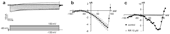
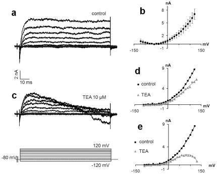
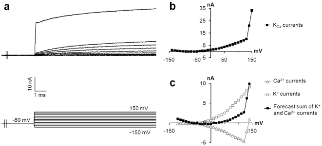
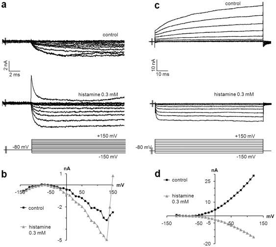
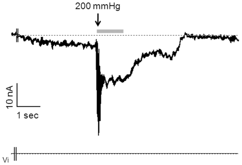
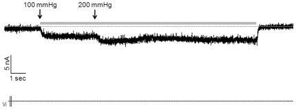
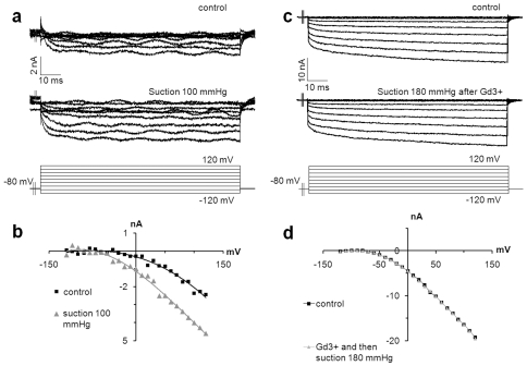
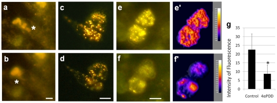
Merkel cells (MCs) associated with nerve terminals constitute MC-neurite complexes, which are involved in slowly-adapting type I mechanoreception. Although MCs are known to express voltage-gated Ca2+ channels and hypotonic-induced membrane deformation is known to lead to Ca2+ transients, whether MCs initiate mechanotransduction is currently unknown. To answer to this question, rat MCs were transfected with a reporter vector, which enabled their identification.Their properties were investigated through electrophysiological studies. Voltage-gated K+, Ca2+ and Ca2+-activated K+ (KCa)channels were identified, as previously described. Here, we also report the activation of Ca2+ channels by histamine and their inhibition by acetylcholine. As a major finding, we demonstrated that direct mechanical stimulations induced strong inward Ca2+ currents in MCs. Depolarizations were dependent on the strength and the length of the stimulation. Moreover, touch-evoked currents were inhibited by the stretch channel antagonist gadolinium. These data confirm the mechanotransduction capabilities of MCs. Furthermore, we found that activation of the osmoreceptor TRPV4 in FM1-43-labeled MCs provoked neurosecretory granule exocytosis. Since FM1-43 blocks mechanosensory channels, this suggests that hypo-osmolarity activates MCs in the absence of mechanotransduction. Thus, mechanotransduction and osmoreception are likely distinct pathways.


Conflict of interest statement

Competing Interests: The authors have declared that no competing interests exist.

Figures

Figure 1
Figure 1. Transfection of MCs from rat vibrissae.
MCs are densely represented in the upper part of the vibrissal outer root sheath as demonstrated by CK20 immunostaining (a). The expression pattern of the neural transcription factor Math1 was restricted to this area, as demonstrated by the read out of the reporter vector pMath1-β-galactosidase (b). Scale bars: 50 µm.
Figure 2
Figure 2. Identification of transfected MCs from rat footpads for electrophysiological analyses.
The epidermis of a rat footpad was stripped off of the dermis and the cells were dissociated and transfected with pMath1-β-galactosidase in order to permit detection of MCs (a). Transfected MCs represented 1.35% (+/−0.73) of the epidermal cells. Electrophysiological recordings targeting blue cells were performed in the cell-attached macro-patch configuration (b). Scale bars: 20 µm.
Figure 3
Figure 3. Voltage-activated Ca2+ channel are produced by MCs.
A majority of MCs expressed Ca2+ currents. (a) Representative inward Ca2+ currents from transfected MCs in response to a series of depolarizing pulses. (b) Average of steady-state current densities during the depolarizing step, plotted as a function of membrane potential. The reverse potential at approximately 150 mV is representative of Ca2+ current (n = 11, error bars are s.e.m.). (c) RR at 10 µM (grey triangles) inhibited inward currents observed in control conditions (black squares), which confirmed the presence of voltage-activated Ca2+ channels.
Figure 4
Figure 4. MCs produced voltage-activated K+ channels.
(a) A representative slow activated outward K+ current lacking an inactivating component. (b) Average steady-state current densities during depolarizing pulses, plotted as a function of voltage (n = 7, error bars denote s.e.m.). (c) TEA (10 mM), a classical inhibitor of K+ channels, decreased current densities compared to control conditions. (a) Non-inactivated currents and slow-activated currents with an inactivating component were observed. Peak (d) and steady-state from the last 40 ms. (e) Current densities during the depolarizing step were plotted as a function of membrane potential for control conditions (black squares) or with TEA (grey triangles). (d) The decreased current densities demonstrated the presence of K+ channels. (e) Steady-state current densities highlighted the inactivating component starting around 80 mV.
Figure 5
Figure 5. Ca2+-activated K+ channels in MCs.
In three recordings, a sharp increase of outward current was detected, suggesting the presence of BKCa channels, which have already been described in MCs. (a) Representative voltage-dependent currents activated in response to a series of depolarizing pulses. (b) The current-voltage curve demonstrated a marked increase of the conductance around +130 mV, suggesting the involvement of different currents. (c) In order to determine whether these currents correspond to both Ca2+ and K+ currents, the two current-voltage curves from a previous recording were added. The pattern of the forecast current to voltage relationship (black squares) corresponded to the recorded currents.
Figure 6
Figure 6. ACh inhibits inward currents in MCs.
(a) Representative inward Ca2+ currents without an inactivating component (top traces) recorded in the control condition from transfected MCs of rat touch dome. These currents were almost entirely inhibited after two min of exposure to ACh (10 µM) (bottom traces). (b) Steady-state current densities were plotted as a function of voltage in normal conditions (black squares) and after stimulation with ACh (grey triangles) (n = 4).
Figure 7
Figure 7. Histamine induces inward currents in MCs.
The histamine H3 receptor was identified at the surface of MCs. Its activation induced inward Ca2+ currents in MCs. Representative current densities recorded during a series of depolarizing steps in control conditions (a, c: top traces). Inward slow activating Ca2+ currents (a) or outward fast activating K+ currents (b) were identified as revealed by the current-voltage relationships (b, d: black squares). In both conditions, the addition of histamine (300 µM) induced inward Ca2+ currents without inactivating components (a, c: bottom traces) as demonstrated by the current-voltage relationships (b, d: grey triangles).
Figure 8
Figure 8. Suction initiates spontaneous inward currents in MCs.
MCs are believed to function as a mechanoreceptors. To determine whether they respond to touch, suction (200 mmHg) was applied to transfected MCs from a rat footpad. Suction induced a strong, non-delayed, inward transient current. The decrease of inward currents at the second part of the recording is explained by the time required to stop the stimulation.
Figure 9
Figure 9. MCs transduce the strength and the length of the stimulation.
A 100 mmHg-suction was applied to transfected MCs. Stimulation induced inward currents sustained throughout the stimulation (about 15 sec). An increase in the strength of the stimulation after 4 sec led to increased inward currents.
Figure 10
Figure 10. Touch-evoked inward currents are inhibited by Gadolinium.
Gd3+ is a stretch-channel inhibitor. Once added to the medium (100 µM), Gd3+ inhibited inward currents induced by suction, supporting the presence of stretch channels in MCs. (a) Representative inward Ca2+ currents recorded in control conditions (top traces) and following the application of suction (bottom traces). (b) Current-voltage relationships demonstrating increased depolarizations during suction (grey triangles) compared to control conditions (black squares). (c) Recorded inward currents in control conditions (top traces) were not modified by suction when cells were pre-exposed to Gd3+ (bottom traces). (d) Steady-state current densities during the depolarizing step were plotted as a function of membrane potential.
Figure 11
Figure 11. FM1–43 stained neurosecretory granules of MCs.
FM1–43 is a fluorescent dye and a permeant blocker of mechanosensory channels, which stained the neurosecretory granules of neurons, hair cells, and MCs. (a) MCs are concentrated in rat footpads as demonstrated by CK20immunostaining. (b) Intraperitoneal injection of FM1–43 at 3 mg/kg in the rat allows for identification of MCs in the basal layer of the epidermis of rat footpads. Scale bars: 50 µm.
Figure 12
Figure 12. TRPV4 agonist induced FM1-43-loaded neurosecretory granule exocytosis.
FM1–43 was injected intraperitoneally (3 mg/kg) in young rats. The following day, MCs from footpads were dissociated and cultured in isotonic medium (a, c, e). Exposure to hypotonic solution (b) or 4αPDD 1 µM (d, f), a TRPV4 agonist, induce neurosecretory granule exocytosis, as suggested by decreases in fluorescence intensity of 61% (n = 33) (g). Computational analyses showed movements of fluorescent particles from the middle to the periphery of the cells (e′, f′) which supported exocytosis. *P<0.01 significantly different from control. Scale bar in all pictures: 5 µm.

References

    1. Lumpkin EA, Caterina MJ. Mechanisms of sensory transduction in the skin. Nature. 2007;445:858–865. - PubMed
    1. Johnson KO. The roles and functions of cutaneous mechanoreceptors. Curr Opin Neurobiol. 2001;11:455–461. - PubMed
    1. Boulais N, Misery L. Merkel cells. J Am Acad Dermatol. 2007;57:147–165. - PubMed
    1. Gaudillere A, Misery L. Merkel cell. Ann Dermatol Venereol. 1994;121:909–917. - PubMed
    1. Mills LR, Diamond J. Merkel cells are not the mechanosensory transducers in the touch dome of the rat. J Neurocytol. 1995;24:117–134. - PubMed

Publication types