

Cardiac conduction system: delineation of anatomic landmarks with multidetector CT
- PMID: 19898655
- PMCID: PMC2766580
Cardiac conduction system: delineation of anatomic landmarks with multidetector CT
Abstract
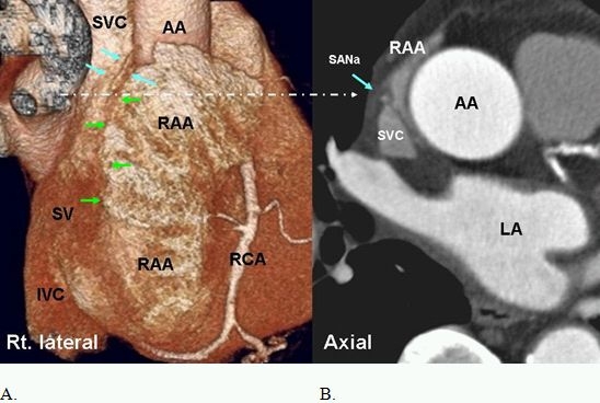
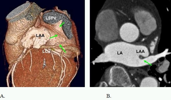
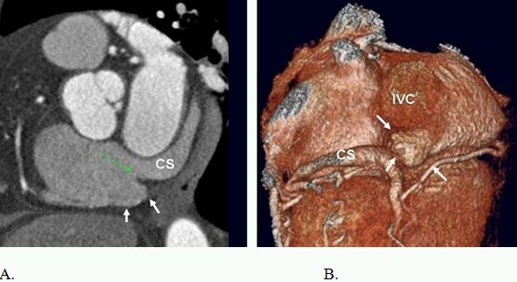
Major components of the cardiac conduction system including the sinoatrial node (SAN), atrioventricular node (AVN), the His Bundle, and the right and left bundle branches are too small to be directly visualized by multidetector CT (MDCT) given the limited spatial resolution of current scanners. However, the related anatomic landmarks and variants of this system a well as the areas with special interest to electrophysiologists can be reliably demonstrated by MDCT. Some of these structures and landmarks include the right SAN artery, right atrial cavotricuspid isthmus, Koch triangle, AVN artery, interatrial muscle bundles, and pulmonary veins. In addition, MDCT has an imperative role in demarcating potential arrhythmogenic structures. The aim of this review will be to assess the extent at which MDCT can outline the described anatomic landmarks and therefore provide crucial information used in clinical practice.
Keywords: Cardiac Conduction System; Delineation of Anatomic Landmarks; Multidetector CT.
Figures


References
-
- Sanchez-Quintana D, et al. Anatomy of cardiac nodes and atrioventricular specialized conduction system. Rev Esp Cardiol. 2003;56:1085. - PubMed
-
- Malouf JF, et al. Functional anatomy of the heart. In: Fuster V, et al., editors. The heart. 11th ed. New York, NY: McGraw-Hill; 2004. p. 75.
-
- Ho SY, et al. Atrial structure and fibres: morphologic bases of atrial conduction. Cardiovasc Res. 2002;54:325. - PubMed
-
- Anderson RH, et al. The anatomy of the heart revisited. Anat Rec. 1996;246:1. - PubMed
-
- Edwards WD. Applied anatomy of the heart. In: Giuliani ER, et al., editors. Cardiology: fundamentals and practice, 2nd ed. St. Louis: St. Louis; 1991. p. 47 .
LinkOut - more resources
Full Text Sources
Other Literature Sources