. 2010 Mar;11(1):39-51.
doi: 10.1007/s10162-009-0196-5. Epub 2009 Nov 7.

Glycosylation regulates prestin cellular activity

Affiliations

Glycosylation regulates prestin cellular activity

Lavanya Rajagopalan et al. J Assoc Res Otolaryngol. 2010 Mar.

Abstract

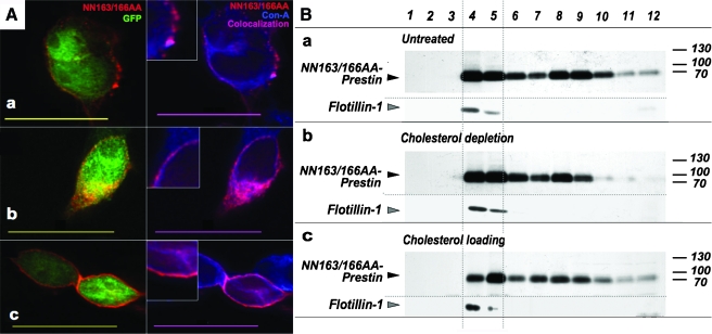
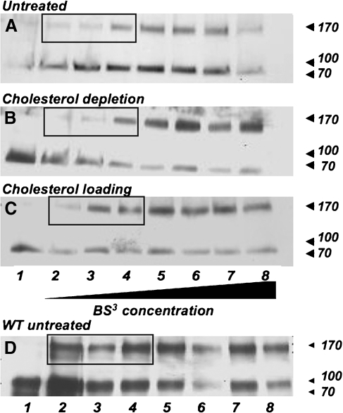
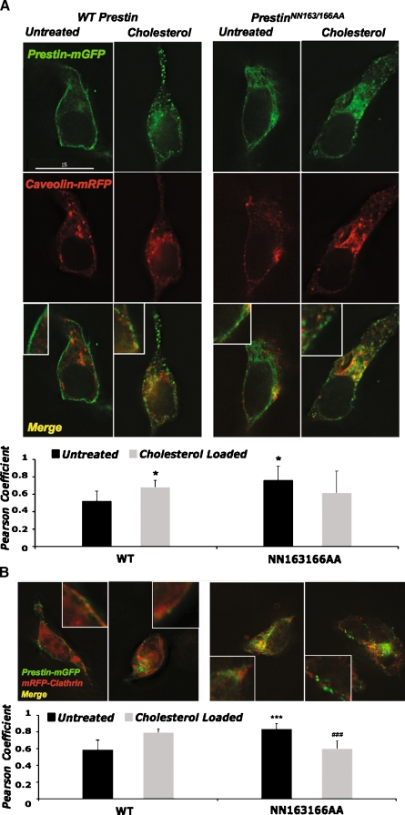
Glycosylation is a common post-translational modification of proteins and is implicated in a variety of cellular functions including protein folding, degradation, sorting and trafficking, and membrane protein recycling. The membrane protein prestin is an essential component of the membrane-based motor driving electromotility changes (electromotility) in the outer hair cell (OHC), a central process in auditory transduction. Prestin was earlier identified to possess two N-glycosylation sites (N163, N166) that, when mutated, marginally affect prestin nonlinear capacitance (NLC) function in cultured cells. Here, we show that the double mutant prestin(NN163/166AA) is not glycosylated and shows the expected NLC properties in the untreated and cholesterol-depleted HEK 293 cell model. In addition, unlike WT prestin that readily forms oligomers, prestin(NN163/166AA) is enriched as monomers and more mobile in the plasma membrane, suggesting that oligomerization of prestin is dependent on glycosylation but is not essential for the generation of NLC in HEK 293 cells. However, in the presence of increased membrane cholesterol, unlike the hyperpolarizing shift in NLC seen with WT prestin, cells expressing prestin(NN163/166AA) exhibit a linear capacitance function. In an attempt to explain this finding, we discovered that both WT prestin and prestin(NN163/166AA) participate in cholesterol-dependent cellular trafficking. In contrast to WT prestin, prestin(NN163/166AA) shows a significant cholesterol-dependent decrease in cell-surface expression, which may explain the loss of NLC function. Based on our observations, we conclude that glycosylation regulates self-association and cellular trafficking of prestin(NN163/166AA). These observations are the first to implicate a regulatory role for cellular trafficking and sorting in prestin function. We speculate that the cholesterol regulation of prestin occurs through localization to and internalization from membrane microdomains by clathrin- and caveolin-dependent mechanisms.


Figures

FIG. 1
FIG. 1
Prestin is a glycoprotein and prestinNN163/166AA is functional. A Prestin is a glycosylated protein, and the prestinNN163/166AA mutant is not glycosylated in HEK 293 cells. Treatment with the glycosidase PNGase-F (to remove N-linked oligosaccharides) alters the gel migration of WT prestin. Untreated WT prestin shows a molecular weight of ∼120 kDa, while treatment with PNGase-F results in a band at ∼80 kDa, corresponding to the mass of deglycosylated prestin. PrestinNN163/166AA mutant forms only one band (at 80 kDa) regardless of PNGase-F treatment. B Cells transfected with NN163/166AA (white circles; n = 8) exhibit a bell-shaped NLC curve with a peak slightly shifted from the WT value of −70 mV (black circles; n = 10). Fit parameters are: for WT, Vpkc = −70 ± 18 mV; valence (z) = 0.82; charge density (Qmax/Clin) = 11 ± 4 fC/pF; for mutant, Vpkc = −55 ± 7.5 mV; valence (z) = 0.84; charge density (Qmax/Clin) = 10.3 ± 3.5 fC/pF. Representative average traces from single cells are shown. Traces have been normalized relative to maximal average NLC of WT. Inset average charge density, calculated from Boltzmann fits to NLC traces, shows no significant differences between WT prestin and prestinNN163/166AA. Error bars represent the SD.
FIG. 2
FIG. 2
Membrane distribution and localization of prestinNN163/166AA. A Immunofluorescence shows prestin fluorescence (red) colocalizes with that of concanavalin A (blue), a membrane marker in all cases. Epifluorescence of GFP (green), which is independently produced from the prestin plasmid as a cytoplasmic protein, has been used to identify transfected cells. a PrestinNN163/166AA exhibits punctate foci of fluorescence in the plasma membrane of HEK 293 cells. b Depletion of cholesterol by MβCD causes a decrease in the size and number of foci. c Loading excess cholesterol causes an apparently homogenous distribution rather than punctate foci of prestinNN163/166AA, consistent with the decreased raft localization see in B, c. Representative images from several (six to eight) cells imaged are shown. B Membrane proteins were fractionated using a sucrose density gradient. a PrestinNN163/166AA is expressed in all membrane fractions, including those containing the raft protein flotillin-1 (lanes 4 and 5). b Depletion of cholesterol does not cause significant redistribution of prestinNN163/166AA in the raft fractions but c loading results in a decrease of prestinNN163/166AA in all fractions, especially in the raft membrane fractions. Shown are representative blots; the experiment has been replicated (n = 3).
FIG. 3
FIG. 3
Cellular cholesterol affects prestinNN163/166AA self-association. A Cross-linking of prestinNN163/166AA in untreated HEK 293 cells with increasing concentrations of the membrane-impermeable agent BS3: higher concentrations of BS3 are required to cross-link the prestinNN163/166AA mutant as oligomers, compared to WT prestin (see boxed lanes, A and D). B Cross-linking of prestinNN163/166AA in cholesterol-depleted HEK 293 cells: similar concentrations of BS3 are required to cross-link prestinNN163/166AA upon cholesterol depletion as in untreated cells (boxed lanes, A and B). C Cross-linking of prestinNN163/166AA in cholesterol-loaded HEK 293 cells: oligomer bands appear at slightly lower levels of cross-linker compared to untreated cells (boxed lanes, A and C). D Cross-linking of WT prestin. In all cases, lanes 1 through 8 represent cross-linking by 0, 0.078, 0.156, 0.312, 0.625, 1.25, 2.5, and 5 mM BS3, respectively. These results are obtained in cells expressing similar levels of WT prestin and prestinNN163/166AA as determined by Western blotting of total prestin (data not shown). Equal loading between lanes was ensured by treating and loading equal aliquots of cells from the same dish. Cross-linking experiments have each been replicated (n = 4); representative blots are shown.
FIG. 4
FIG. 4
Cholesterol affects membrane capacitance and peak voltage of HEK 293 cells expressing prestinNN163/166AA. A Untreated cells expressing prestinNN163/166AA exhibit a bell-shaped NLC function (black squares; n = 8). Fit parameters: Vpkc = −55 ± 7.5 mV; valence (z) = 0.84; charge density (Qmax/Clin) = 10.3 ± 3.5 fC/pF. The NLC peak shifts towards depolarized voltages, similar to the WT trend (Sturm et al. ; Rajagopalan et al. 2007), in cells depleted of cholesterol (white squares; n = 6) in comparison to untreated cells. Fit parameters: Vpkc = −0.16 ± 8.9 mV; valence (z) = 0.81; charge density (Qmax/Clin) = 7.4 ± 9.3 fC/pF. However, cells loaded with cholesterol exhibit loss of the bell-shaped capacitance curve and exhibit linear capacitance (multiplication sign; n = 6) similar to untransfected and mock-transfected HEK 293 cells. Representative average traces from single cells are shown. Traces have been normalized relative to baseline and peak voltage. B Mean voltage at peak capacitance (Vpkc) for WT prestin and prestinNN163/166AA in HEK 293 cells. All parameters are similar between WT and prestinNN163/166AA, except in the case of cholesterol loading. Error bars represent SD. The gray box represents the normal range of Vpkc for WT prestin. C Change in Vpkc in representative single cells transfected with WT (black circles) or prestinNN163/166AA (white circles) upon cholesterol loading. D Change in Vpkc in representative single cells transfected with WT (black circles) or prestinNN163/166AA (white circles) upon cholesterol loading (black arrow) followed by depletion (gray arrows). E Change in charge density in representative single cells transfected with WT (black circles) or prestinNN163/166AA (white circles) upon cholesterol loading. Cells transfected with prestinNN163/166AA exhibited an average charge density reduction of 33.8 ± 5.9% from initial values 20 min after the addition of cholesterol, compared with no significant change in WT. F Change in linear capacitance in representative single cells transfected with WT (black circles) or prestinNN163/166AA (white circles) upon cholesterol loading. In CF, the black arrow indicates time of loading and the gray arrow indicates the time of depletion using MβCD. Data from representative single cells are shown; the experiments have been repeated in four cells for each treatment.
FIG. 5
FIG. 5
WT prestin and prestinNN163/166AA colocalize with caveolin and clathrin vesicles. Atop deconvolution images of WT prestin-mGFP and prestinNN163/166AA-mGFP in HEK 293 cells show colocalization of prestin (green) with Cav1-mRFP (red); bottom the Pearson's coefficient of correlation between prestin and caveolin fluorescence indicates a significant increase in caveolin colocalization with WT prestin-mGFP upon cholesterol loading. The prestinNN163/166AA-mGFP, which colocalizes more strongly than WT in untreated cells, does not show significant differences in colocalization upon cholesterol loading. *p < 0.05, statistical significance in comparison to WT untreated as determined by Student's t tests. Data represent the mean ± SD from five to seven cells per group. Btop deconvolution images of WT prestin-mGFP and prestinNN163/166AA-mGFP in HEK cells show colocalization of prestin (green) with mRFP-clathrin (red); bottom the Pearson's coefficient of correlation between prestin and clathrin fluorescence indicates a significant increase in clathrin colocalization with WT prestin-mGFP upon cholesterol loading. PrestinNN163/166AA-mGFP colocalizes more strongly with clathrin than WT in untreated cells, but this colocalization decreases significantly upon cholesterol loading. **p < 0.01; ***p < 0.005, statistical significance (determined by Student's t tests; comparison to WT untreated) or ###p < 0.005, statistical significance (determined by Student's t tests; comparison to prestinNN163/166AA untreated). Data represent the mean ± SD from five to six cells per group.
FIG. 6
FIG. 6
PrestinNN163/166AA has higher membrane mobility; cholesterol loading causes a decrease in mobility and cell-surface population. A The relative diffusion of NN163/166AA prestin is significantly higher than that of WT. Cholesterol loading has a minimal effect of the lateral diffusion of WT prestin, but dramatically reduced the lateral diffusion of mutant prestin. All diffusion measurements have been normalized to that of WT. B In the first bleach, both constructs show an increase in IF upon loading, indicative of an increase in endocytotic vesicles/caveolae. However, with this first IF effectively discounted by the first bleach, the second bleach shows no change in the IF of WT prestin upon cholesterol loading, while mutant prestin exhibits a large increase in IF. Values are shown as the mean ± SD. *p < 0.05, **p < 0.01, ***p < 0.005; significance was determined using analysis of variance with Tukey's honestly significant difference test for diffusion measurements and Student's t tests for IF data. For A and B, sample sizes are as follows: WT untreated, n = 21; WT loaded, n = 10; mutant untreated, n = 12; mutant loaded, n = 9. C The relative cell-surface population of WT prestin after cholesterol loading (compared to untreated cells) is unchanged on average, while prestinNN163/166AA shows a significant (>40%; p < 0.05) decrease in cell-surface population in cholesterol-loaded cells when compared to untreated cells. Plotted are average relative cell-surface populations of cholesterol-loaded HEK 293 cells (compared to untreated cells expressing the same prestin variant), calculated from three separate experiments. Comparisons between WT prestin and prestinNN163/166AA have not been made due to intrinsic expression differences between the WT and mutant plasmids. D Representative blots of WT and prestinNN163/166AA cell-surface populations in untreated and cholesterol-loaded cells. Protein concentrations were determined for each sample in order to load equal amounts of whole-cell lysates for fractionation.

References

    1. Adler HJ, Belyantseva IA, Merritt RCJ, Frolenkov GI, Dougherty GW, Kachar B. Expression of prestin, a membrane motor protein, in the mammalian auditory and vestibular periphery. Hear Res. 2003;184:27–40. doi: 10.1016/S0378-5955(03)00192-8. - DOI - PubMed
    1. Ashmore J. Cochlear outer hair cell motility. Physiol Rev. 2008;88:173–210. doi: 10.1152/physrev.00044.2006. - DOI - PubMed
    1. Belyantseva IA, Adler HJ, Curi R, Frolenkov GI, Kachar B. Expression and localization of prestin and the sugar transporter GLUT-5 during development of electromotility in cochlear outer hair cells. J Neurosci. 2000;20:RC116. - PMC - PubMed
    1. Brownell WE. The piezoelectric hair cell. In: Eatock RA, editor. Vertebrate hair cells. New York: Springer; 2006. pp. 313–347.
    1. Brownell WE, Oghalai JS. Structural basis of outer hair cell motility or where's the motor? In: Lim D, editor. Cell and molecular biology of the ear. New York: Academic; 2000. pp. 69–83.

Publication types

MeSH terms

LinkOut - more resources