. 2010 May;15(3):281-93.
doi: 10.1007/s12192-009-0142-9. Epub 2009 Nov 8.

Plasma cell differentiation initiates a limited ER stress response by specifically suppressing the PERK-dependent branch of the unfolded protein response

Affiliations

Plasma cell differentiation initiates a limited ER stress response by specifically suppressing the PERK-dependent branch of the unfolded protein response

Yanjun Ma et al. Cell Stress Chaperones. 2010 May.

Abstract

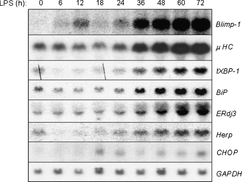
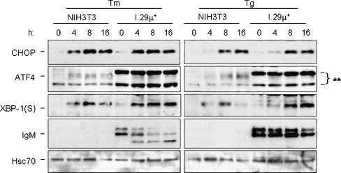
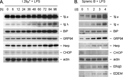
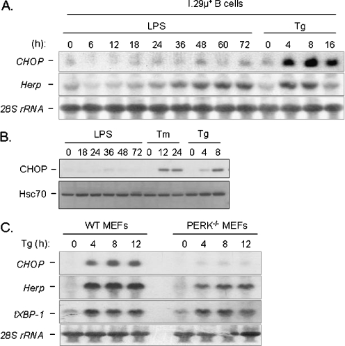
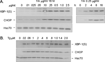
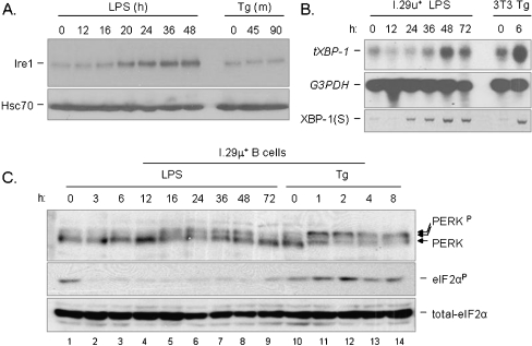
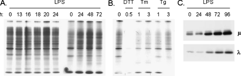
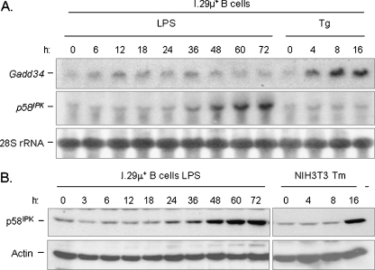
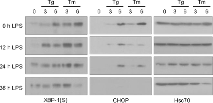
In response to terminal differentiation signals that enable B cells to produce vast quantities of antibodies, a dramatic expansion of the secretory pathway and a corresponding increase in the molecular chaperones and folding enzymes that aid and monitor immunoglobulin synthesis occurs. Recent studies reveal that the unfolded protein response (UPR), which is normally activated by endoplasmic reticulum (ER) stress, plays a critical role in this process. Although B cells activate all three branches of the UPR in response to pharmacological inducers of the pathway, plasma cell differentiation elicits only a partial UPR in which components of the PKR-like ER kinase (PERK) branch are not expressed. This prompted us to further characterize UPR activation during plasma cell differentiation. We found that in response to lipopolysaccharides (LPS)-induced differentiation of the I.29 micro(+) B cell line, Ire1 was activated early, which led to splicing of XBP-1. PERK was partially phosphorylated with similar kinetics, but this was not sufficient to activate its downstream target eIF-2alpha, which initiates translation arrest, or to induce other targets like CHOP or GADD34. Both of these events preceded increased Ig synthesis, arguing this is not the signal for activating these two transducers. Targets of activating transcription factor 6 (ATF6) were up-regulated considerably later, arguing that the ATF6 branch is activated by a distinct signal. Pretreatment with LPS inhibited activation of the PERK branch by pharmacological inducers of the UPR, suggesting that differentiation-induced signals specifically silence this branch. This unique ability to differentially regulate various branches of the UPR allows B cells to accomplish distinct outcomes via the same UPR machinery.


Figures

Fig. 1
Fig. 1
UPR targets are activated during the differentiation of I.29 μ+ B cells into plasma cells upon LPS stimulation. Cytosolic RNA from I.29 μ+ cells treated with LPS for the indicated periods of time were electrophoresed, transferred, and then probed with plasma cell markers like Blimp-1 and μ heavy chain (μ HC) or UPR targets like XBP-1, BiP, ERdj3, Herp, and CHOP. GAPDH was probed as control for loading
Fig. 2
Fig. 2
The Ire1, ATF6, and PERK branches of the UPR are intact in I.29 μ+ cells. NIH3T3 fibroblasts or I.29 μ+ B cells were treated with the ER stressors tunicamycin (Tm) or thapsigargin (Tg) for the indicated period of time. Cell lysates were harvested and separated on 10% SDS-polyacrylamide gels and transferred for western blot analyses. The membrane was then blotted for the PERK targets CHOP and ATF4, the Ire1/ATF6 regulated target, XBP-1(S), and mouse μ heavy chain (which is only present in I.29 μ+ cells). Hsc70 was examined as a control for loading. The asterisks indicate two non-specific bands detected with the ATF4 antibody in I.29 μ+ cells
Fig. 3
Fig. 3
Splenic B cells show similar induction of UPR targets with LPS as I.29 μ+ cells as well as the differential induction of CHOP and Herp proteins. Cell lysates from I.29 µ+ B cells (a) or mouse splenic B cells (b) were treated with LPS for the indicated times and equal amounts of protein were loaded for each sample. After separation by SDS-PAGE and transfer, membranes were probed with the indicated antibodies. Actin served as a control for loading
Fig. 4
Fig. 4
UPR targets CHOP and Herp are regulated differently during B-cell differentiation. a Cytosolic RNA from I.29 µ+ B cells stimulated with either LPS or Tg for the indicated periods of time were harvested, separated, and then probed for CHOP and Herp. 28S rRNA was probed as a loading control. b Cell extracts were prepared from cells treated as indicated and CHOP protein expression was determined by Western blotting. Hsc70 protein served as a control for loading. c Cytosolic RNA from WT and PERK null MEFs treated with Tg for the indicated periods of time were harvested, separated, and then probed for CHOP, Herp, and total XBP-1 (tXBP-1) transcript levels. Again, 28S rRNA was probed as a loading control
Fig. 5
Fig. 5
Ire1 and PERK are equally sensitive to ER stress induced by conventional stressors like Tm or Tg. a Cell lysates from I.29 μ+ B cells treated for 16 h with the indicated concentrations of Tm (left panel) or 0.25 ug/ml of Tm for the indicated periods of time (right panel) were harvested, separated on 10% SDS-polyacrylamide gel and transferred to a PVDF membrane. The membrane was then blotted for CHOP and XBP-1(S). Hsc70 was examined as a loading control. b Cell lysates were prepared from I.29 μ+ B cells that had been treated for 16 h with the indicated concentrations of Tg and examined as in a
Fig. 6
Fig. 6
Both Ire1 and PERK are activated to different extents during plasma cell differentiation, but eIF-2α phosphorylation downstream of PERK is blocked. a. Cell lysates from I.29 μ+ B cells treated with either LPS or Tg for the indicated period time was harvested and then separated on a 10% SDS-PAGE. The membrane was blotted for Ire1 and for Hsc70 as a loading control. b Cytosolic RNA from I.29 μ+ cells treated with LPS, or NIH3T3 cells treated with Tg for the indicated period of time was harvested and separated. Total XBP-1 (tXBP-1) and GAPDH mRNA levels were then probed, the latter as a loading control. RT-PCR was also performed with the same RNA samples using primers specific for the spliced form of XBP-1 (sXBP-1). c Cell lysates from I.29 μ+ cells treated with LPS or Tg for the indicated period of time were separated on a 8% SDS-PAGE which was then blotted for PERK, total and phosphorylated form of eIF-2α
Fig. 7
Fig. 7
LPS induced differentiation does not induce an arrest in protein synthesis. I.29 μ+ B cells were treated with LPS (a) or conventional UPR inducers (b) for the indicated times and then pulse-labeled with [35S] methionine and cysteine for 5 min. Cell lysates were analyzed directly by SDS-PAGE and proteins were visualized by autoradiography. c An aliquot of cell lysates from the indicated times after LPS induction were examined by Western blotting for Ig heavy and light chain expression
Fig. 8
Fig. 8
p58IPK, but not GADD34, is induced during the differentiation of I.29 μ+ B cells into plasma cells. a I.29 μ+ B cells were treated with LPS or Tg for the indicated periods of time and RNA was isolated and prepared for Northern blotting. The membrane was probed for GADD34 and p58IPK. 28S rRNA levels were also examined to control for loading. b Cell lysates were prepared from I.29 μ+ B cells that had been treated with LPS or from NIH3T3 fibroblasts treated with Tm for the indicated periods of time. Samples were examined on a 10% SDS-PAGE and transferred. The membrane was then blotted for p58IPK protein and actin as a loading control
Fig. 9
Fig. 9
LPS pretreatment of I.29 μ+ B cells augment XBP-1(S) levels but inhibits CHOP induction by pharmacological ER stressors. I.29 μ+ B cells were pretreated with LPS for 0, 12, 24, or 36 h and then further cultured with Tm or Tg for 0, 3 or 6 h. Cell lysates were harvested for each experimental group and analyzed by SDS-PAGE. After transferring, the membranes were blotted for XBP-1(S) (left panel), CHOP (middle panel) or Hsc70 (right panel) as a control for loading

Similar articles

Cited by

References

    1. Bertolotti A, Zhang Y, Hendershot LM, Harding HP, Ron D. Dynamic interaction of BiP and ER stress transducers in the unfolded-protein response. Nat Cell Biol. 2000;2:326–332. doi: 10.1038/35014014. - DOI - PubMed
    1. Brewer JW, Diehl JA. PERK mediates cell-cycle exit during the mammalian unfolded protein response. Proc Natl Acad Sci USA. 2000;97:12625–12630. doi: 10.1073/pnas.220247197. - DOI - PMC - PubMed
    1. Brewer JW, Cleveland JL, Hendershot LM. A pathway distinct from the mammalian unfolded protein response regulates expression of endoplasmic reticulum chaperones in non-stressed cells. EMBO J. 1997;16:7207–7216. doi: 10.1093/emboj/16.23.7207. - DOI - PMC - PubMed
    1. Calfon M, Zeng H, Urano F, Till JH, Hubbard SR, Harding HP, Clask SG, Ron D. IRE1 couples endoplasmic reticulum load to secretory capacity by processing the XBP-1 mRNA. Nature. 2002;415:92–96. doi: 10.1038/415092a. - DOI - PubMed
    1. Gass JN, Gifford NM, Brewer JW. Activation of an unfolded protein response during differentiation of antibody-secreting B cells. J Biol Chem. 2002;277:49047–49054. doi: 10.1074/jbc.M205011200. - DOI - PubMed

Publication types

MeSH terms