Review
. 2010 Feb;216(2):209-22.
doi: 10.1111/j.1469-7580.2009.01157.x. Epub 2009 Nov 9.

The evolution of anatomical illustration and wax modelling in Italy from the 16th to early 19th centuries

Affiliations
Review

The evolution of anatomical illustration and wax modelling in Italy from the 16th to early 19th centuries

Alessandro Riva et al. J Anat. 2010 Feb.

Abstract

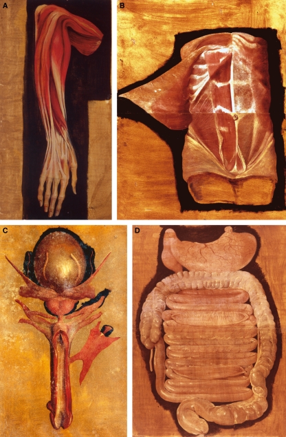
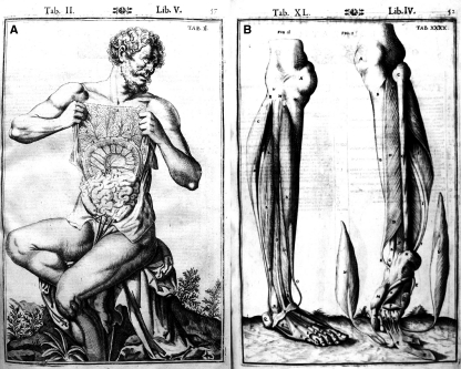
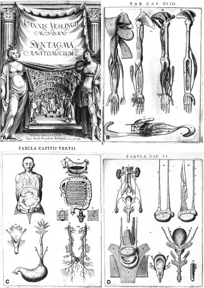
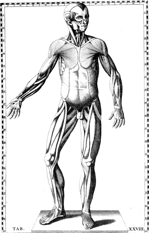
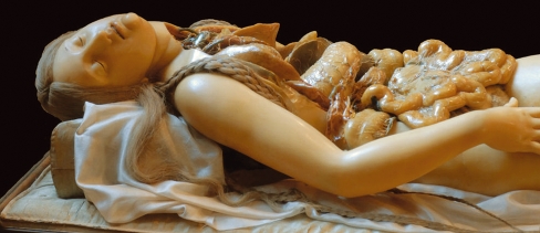
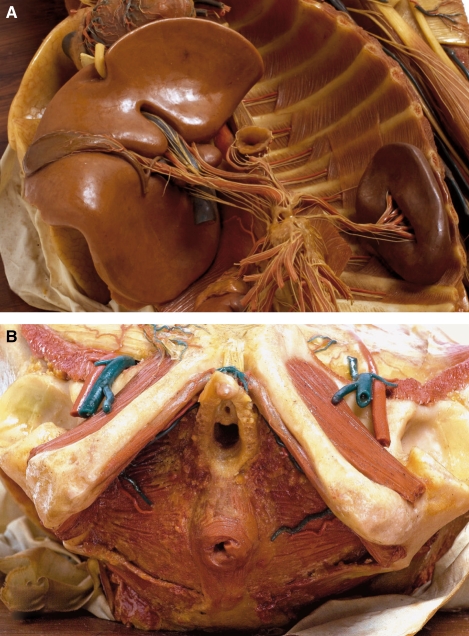
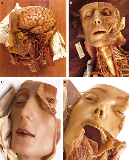
Although the contribution to anatomical illustration by Vesalius and his followers has received much attention, less credit has been given to Veslingius and particularly Fabricius. By 1600, Fabricius had amassed more than 300 paintings that together made the Tabulae Pictae, a great atlas of anatomy that was highly admired by his contemporaries. Many of his new observations were incorporated into subsequent books, including those by Casserius, Spighelius, Harvey and Veslingius. Also of importance were the Tabulae by Eustachius (1552), which, although only published in 1714, greatly influenced anatomical wax modelling. In 1742, Pope Benedict XIV established a Museum of Anatomy in Bologna, entrusting to Ercole Lelli the creation of several anatomical preparations in wax. Felice Fontana realised that the production of a large number of models by the casting method would make cadaveric specimens superfluous for anatomical teaching and in 1771 he asked the Grand Duke to fund a wax-modelling workshop in Florence as part of the Natural History Museum, later known as La Specola. Fontana engaged Giuseppe Ferrini as his first modeller and then the 19-year-old Clemente Susini who, by his death in 1814, had superintended the production of, or personally made, more than 2000 models. In 1780, the Austrian Emperor Joseph II visited La Specola and ordered a great number of models for his Josephinum museum; these were made by Fontana with the help of Clemente Susini and supervised by the anatomist Paolo Mascagni. It is, however, in Cagliari that some of Susini's greatest waxes are to be found. These were made when he was free of Fontana's influence and were based on dissections made by Francesco Antonio Boi (University of Cagliari). Their distinctive anatomical features include the emphasis given to nerves and the absence of lymphatics in the brain, a mistake made on earlier waxes. The refined technical perfection of the anatomical details demonstrates the closeness of the cooperation between Susini and Boi, whereas the expressiveness of the faces and the harmony of colours make the models of Cagliari masterpieces of figurative art.


Figures

Fig. 1
Fig. 1
Fabricius: Tabulae Pictae. (A) Rari 116.16 (De anatomia muscolorum totius corporis): muscles of the right forearm; (B) Rari 117 1-2 (De anatomia): abdominal muscles and fasciae; (C) Rari 117.23 (De anatomia): male genital apparatus; (D) Rari 117.11–12 (De anatomia): small and large intestines, with demonstration of the rugae. Courtesy of the Marciana Library, Venice.
Fig. 2
Fig. 2
Casserius: Tabulae Anatomicae (1627). (A) Tab. II Lib. V: the figure, holding his own omentum, is standing in a beautiful garden; (B) Tab. XL Lib. IV: the dissected legs are standing on a lawn. Courtesy of Biblioteca Comunale Bonetta, Pavia.
Fig. 3
Fig. 3
Johannes Vesling: Syntagma Anatomicum (1647). (A) Frontispiece: Vesling is depicted during a dissection held in Fabricius’s anatomical theatre in Padua; (B) Tab. Cap. XVIII: muscles of right upper arm, and of the left hand; (C) Tab. Cap. III: organs of the digestive tract and diagram of visceral nerves; (D) Tab. Cap. VI: male urogenital system. (A) Courtesy of Biblioteca Pinali Antica, Padua. (B–D) Courtesy of Thomas Fisher Rare Book Library, University of Toronto.
Fig. 4
Fig. 4
Eustachius: Tabulae Anatomicae (1714): Tab. XVIII, muscular figure. Courtesy of Biblioteca Centrale Biomedica, Cagliari.
Fig. 5
Fig. 5
Detail of the statue representing ‘deep lymphatic vessels in a female subject’, ordered from Fontana by Scarpa and executed by Susini in 1794. Note the sleeping, serene expression of the face, typical of Fontana’s manner. Museum of the History of the University, Pavia. Photography by Alberto Calligaro.
Fig. 6
Fig. 6
Fresco commissioned by Giovanni Alessandro Brambilla in the ceiling of Pavia anatomical theatre (now Aula Scarpa) to celebrate the newly achieved parity between Surgery and Medicine. Photography by Alberto Calligaro.
Fig. 7
Fig. 7
Exhibition Room of the Museum of Cagliari. Photography by Gabriele Conti.
Fig. 8
Fig. 8
Portrait of Francesco Antonio Boi. Unknown painter of the second half of the 19th century. Photography by Alessandro Cadau.
Fig. 9
Fig. 9
Examples from the Cagliari anatomical wax collection. (A) Detail of case XII model showing the coeliac plexus; note that the hepatic artery is double. (B) Detail of case III model: dissection of the female perineum. Note the relationship of the bulbs of the vestibule (officially renamed corpora spongiosa clitoridis by the Federative International Committee on Anatomical Terminology, 2007) with the urethra and clitoris. Photography by Dessì & Monari.
Fig. 10
Fig. 10
Cagliari collection. (A) Detail of case XIII model: the brain is devoid of lymphatics; note also the detailed representation of nerves and the correct configuration of cerebral convolutions. (B) Detail of case XII model; note the tag with Susini’s signature and date. (C) Detail of case XVIII model. (D) Detail of case XVI model. Photography by Dessì and Monari.
Fig. 11
Fig. 11
Advertising poster based on model III of the Cagliari Collection. Spectacular Bodies (curators: M. Kemp and M. Wallace), Hayward Gallery, London, 19 October 2001–14 January 2002. Concept by Publicis. Photography by Masuad Golsorkhi.

Similar articles

Cited by

References

    1. Azzaroli ML. La ceroplastica nella scienza e nell’arte. Atti del 1 Congresso Internazionale. Firenze: Leo S Olschki Editore; 1977. La Specola. The zoological museum of Florence University; pp. 1–22.
    1. Ballestriero R. The art of ceroplastics. Clemente Susini and the collection of the anatomical wax models of the University of Cagliari. In: Riva A, editor. Flesh and Wax. The Clemente Susini’s Anatomical Models in the University of Cagliari. Nuoro: Ilisso; 2007. pp. 35–45.
    1. Ballestriero R. Anatomical models and wax venuses: art masterpieces or scientific craft works? J Anat. 2010;216:223–234. - PMC - PubMed
    1. Bates AW. ‘Indecent and demoralising representations’: public anatomy museums in mid-Victorian England. Med Hist. 2008;52:1–22. - PMC - PubMed
    1. Belloni L. Per la storia della medicina. Bologna: Arnaldo Forni Editore; 1980.