. 2010 Jan;216(1):23-36.
doi: 10.1111/j.1469-7580.2009.01161.x. Epub 2009 Nov 9.

Functional morphology of the sound-generating labia in the syrinx of two songbird species

Affiliations

Functional morphology of the sound-generating labia in the syrinx of two songbird species

Tobias Riede et al. J Anat. 2010 Jan.

Abstract

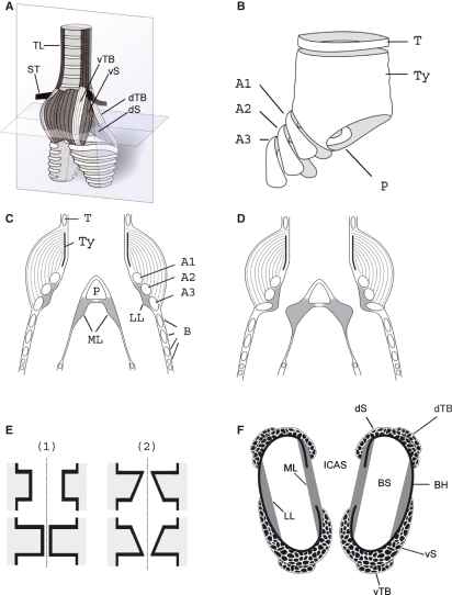
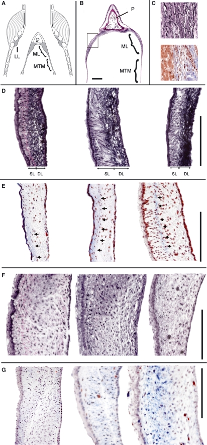
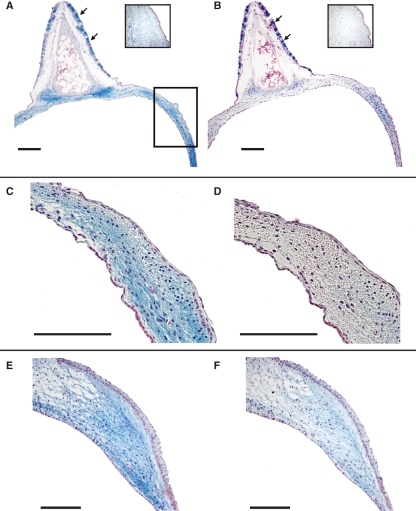
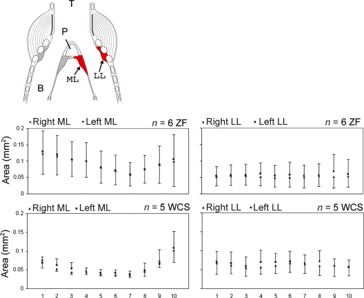
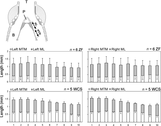
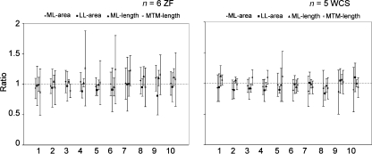
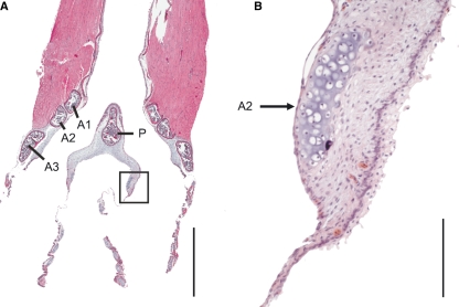
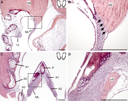
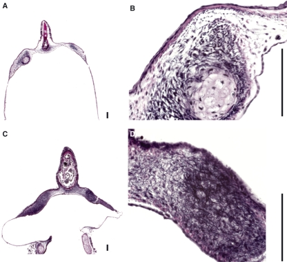
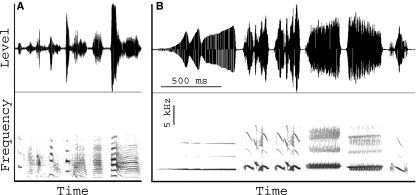
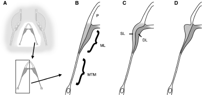
In songbirds, two sound sources inside the syrinx are used to produce the primary sound. Laterally positioned labia are passively set into vibration, thus interrupting a passing air stream. Together with subsyringeal pressure, the size and tension of the labia determine the spectral characteristics of the primary sound. Very little is known about how the histological composition and morphology of the labia affect their function as sound generators. Here we related the size and microstructure of the labia to their acoustic function in two songbird species with different acoustic characteristics, the white-crowned sparrow and zebra finch. Histological serial sections of the syrinx and different staining techniques were used to identify collagen, elastin and hyaluronan as extracellular matrix components. The distribution and orientation of elastic fibers indicated that the labia in white-crowned sparrows are multi-layered structures, whereas they are more uniformly structured in the zebra finch. Collagen and hyaluronan were evenly distributed in both species. A multi-layered composition could give rise to complex viscoelastic properties of each sound source. We also measured labia size. Variability was found along the dorso-ventral axis in both species. Lateral asymmetry was identified in some individuals but not consistently at the species level. Different size between the left and right sound sources could provide a morphological basis for the acoustic specialization of each sound generator, but only in some individuals. The inconsistency of its presence requires the investigation of alternative explanations, e.g. differences in viscoelastic properties of the labia of the left and right syrinx. Furthermore, we identified attachments of syringeal muscles to the labia as well as to bronchial half rings and suggest a mechanism for their biomechanical function.


Figures

Fig. 1
Fig. 1
The songbird syrinx. (A) Schematic external ventral view of the excised organ. TL, tracheolateralis muscle; ST, sternotrachealis muscle; dTB and vTB, dorsal and ventral tracheobronchial muscle; dS and vS, dorsal and ventral syringeal muscle. (B) Schematic view of the cartilage framework of the syrinx (modified after Ames, 1971). The drum [or tympanum (Ty)] is located at the caudal end of the trachea. Its shape varies with species. A median dorso-ventral bar [pessulus (P)], spanning the lumen of the trachea, marks the end of the trachea and the beginning of the primary bronchi. The pessulus is one important attachment for the ML. A1–A3, bronchial half rings; T, tracheal ring. (C) Schematic of a frontal section of the syrinx (level indicated by the vertical plate in A). B, bronchial rings. (D) Schematic of the adduction setting of the ML and LL. (E) Simulating the labia oscillation assumes two major movement components: (1) a latero-lateral component and (2) a cranio-caudal component. The latter leads to a repeated change between a convergent and divergent cross-sectional profile. This out-of-phase movement of the upper and lower part of the labia produces larger asymmetries in the forces acting on the labia in the opening and closing phase of the oscillation than the inward and outward movement alone, resulting in a positive pressure on the labia during the opening phase and a net energy input to the labia maintaining a self-sustained oscillation. (F) Schematic horizontal section through the syrinx (level indicated by the horizontal plate in A). BS, bronchial lumen; BH, bronchial half ring.
Fig. 2
Fig. 2
Elastica van Gieson (EVG) and trichrome (TRI) stain. (A) Schematic of the syrinx frontal section. (B) Section of the pessulus, ML and MTM. The square indicates the location of the higher magnification images in D–G. (C) Control stains for EVG (upper square) and TRI (lower square). In D–G, the tracheal lumen is to the left and the air sac lumen is to the right. (D) EVG stain in three specimens of the WCS. Note that most of the elastic fibers in the WCS are concentrated near the air sac side of the labium. (E) Masson’s TRI stain in three specimens of the WCS. The arrows indicate a local concentration of blue-stained collagen fibers. (F) EVG stain in three specimens of the ZF. (G) Masson’s TRI stain in three specimens of the ZF. P, pessulus; SL, superficial layer; DL, deep layer. Bar: 100 μm.
Fig. 3
Fig. 3
ML. (A,C,E) Alcian blue stain. (B,D,F) Hyaluronidase digestion and Alcian blue stain, same specimens as in A,C,E, respectively. (A–D) WCS. (E and F) ZF. The insets in A and B are control stains for Alcian blue stain and hyaluronidase digestion and Alcian blue stain. The square in A indicates the location of the higher magnification images in C–F. Goblet cells, located in the ciliated epithelium of the semilunar membrane, are indicated by arrows. Bars: 100 μm.
Fig. 5
Fig. 5
Cross-sectional area measurements of the ML and LL. Squares (left side) and triangles (right side) indicate means and error bars indicate SD. T, trachea; B, bronchus; P, pessulus.
Fig. 4
Fig. 4
Cranio-caudal length of the ML and MTM. Bars indicate means and error bars indicate SD. Gray bar, ML; open bar, MTM; T, trachea; B, bronchus; P, pessulus.
Fig. 6
Fig. 6
Ratios between the right and left syrinx for four measurements (ML area, LL area, ML length and MTM length; Mean ± SD). Horizontal dotted lines indicates a ratio of 1.
Fig. 7
Fig. 7
Frontal section from a ventral aspect of a ZF syrinx. The bronchial half ring arches into the ML ventrally and dorsally (see also Fig. 1F). The square in A indicates the area of magnification in B. A1, A2 and A3, first, second and third bronchial half ring, P, pessulus. Bars: in A: 1 mm; in B: 100 μm.
Fig. 8
Fig. 8
Two sections from a WCS syrinx indicating direct muscle attachments on labial tissue. The insets at the top right corners of A and C indicate the sectioning level along the dotted line (compare Fig. 1F). (A) Section through the dorsal part of the syrinx. The square in A indicates the area of magnification in B. Note that some fibers of the dorsal syringeal muscle (dS) attach directly to the ML (indicated by arrows). (C) Section through the ventral part of the syrinx. (D) Magnification of the medial aspect of the ML (as indicated by a square in C). The ventral syringeal muscle (vS) attaches to the ML. A1, A2 and A3, first, second and third bronchial half ring; P, pessulus. Bars: 100 μm.
Fig. 9
Fig. 9
The area around the endpoints of the bronchial half rings shows many elastic fibers indicating that the connection between cartilage and soft tissue is enhanced/stabilized, suggestive of a permanent force acting there. (A,B) WCS; (C,D) ZF. P, pessulus. Bars: 100 μm.
Fig. 10
Fig. 10
Waveforms (upper panel) and spectrograms (lower panel) of songs from a male ZF (A) and a male WCS (B). Units in B apply to both panels. Horizontal lines and numbers in the spectrogram indicate the duration of individual acoustic units (‘syllables’).
Fig. 11
Fig. 11
Schematic frontal section of the syrinx illustrating recruitment of layers into oscillation. The details in B–D are indicated in A (compare Fig. 1C,D). The two layers of the ML can be recruited variably into oscillation. (B) Relaxed position of the ML. (C) The ML is uniformly deformed. (D) The superficial layer is selectively involved in oscillation. P, pessulus; SL, superficial layer; DL, deep layer.

References

    1. Amador A, Goller F, Mindlin GB. Frequency modulation during song in a suboscine does not require vocal muscles. J Neurophysiol. 2008;99:2383–2389. - PubMed
    1. Ames PL. The morphology of the syrinx in passerine birds. New Haven, Connecticut: Peabody Museum of Natural History, Yale University; 1971.
    1. Burke MR, Adkins-Regan E, Wade J. Laterality in syrinx muscle morphology of the Japanese quail (Coturnix japonica) Physiol Behav. 2007;90:682–686. - PMC - PubMed
    1. Caton TBS, Thibeault SL, Klemuk S, et al. Viscoelasticity of hyaluronan and nonhyaluronan based vocal fold injectables: implications for mucosal versus muscle use. Laryngoscope. 2007;117:516–521. - PubMed
    1. Chamberlain DR, Gross WB, Cornwell GW, et al. Syringeal anatomy in the common crow. Auk. 1968;85:244–252.

Publication types