. 2010 Jan 22;395(3):544-57.
doi: 10.1016/j.jmb.2009.11.001. Epub 2009 Nov 6.

F-actin structure destabilization and DNase I binding loop: fluctuations mutational cross-linking and electron microscopy analysis of loop states and effects on F-actin

Affiliations

F-actin structure destabilization and DNase I binding loop: fluctuations mutational cross-linking and electron microscopy analysis of loop states and effects on F-actin

Zeynep A Oztug Durer et al. J Mol Biol. .

Abstract

The conformational dynamics of filamentous actin (F-actin) is essential for the regulation and functions of cellular actin networks. The main contribution to F-actin dynamics and its multiple conformational states arises from the mobility and flexibility of the DNase I binding loop (D-loop; residues 40-50) on subdomain 2. Therefore, we explored the structural constraints on D-loop plasticity at the F-actin interprotomer space by probing its dynamic interactions with the hydrophobic loop (H-loop), the C-terminus, and the W-loop via mutational disulfide cross-linking. To this end, residues of the D-loop were mutated to cysteines on yeast actin with a C374A background. These mutants showed no major changes in their polymerization and nucleotide exchange properties compared to wild-type actin. Copper-catalyzed disulfide cross-linking was investigated in equimolar copolymers of cysteine mutants from the D-loop with either wild-type (C374) actin or mutant S265C/C374A (on the H-loop) or mutant F169C/C374A (on the W-loop). Remarkably, all tested residues of the D-loop could be cross-linked to residues 374, 265, and 169 by disulfide bonds, demonstrating the plasticity of the interprotomer region. However, each cross-link resulted in different effects on the filament structure, as detected by electron microscopy and light-scattering measurements. Disulfide cross-linking in the longitudinal orientation produced mostly no visible changes in filament morphology, whereas the cross-linking of D-loop residues >45 to the H-loop, in the lateral direction, resulted in filament disruption and the presence of amorphous aggregates on electron microscopy images. A similar aggregation was also observed upon cross-linking the residues of the D-loop (>41) to residue 169. The effects of disulfide cross-links on F-actin stability were only partially accounted for by the simulations of current F-actin models. Thus, our results present evidence for the high level of conformational plasticity in the interprotomer space and document the link between D-loop interactions and F-actin stability.


Figures

Figure 1
Figure 1
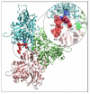
A three protomer Oda model of the actin filament shown with the cross-linked regions highlighted. Residues of the D-loop are colored red, the C-terminal residue C374 is colored blue, the W-loop residue Y169 (mutated to cysteine in this study) is colored blue, and the H-loop residue S265 is colored green (mutated to cysteine in this study). Individual protomers are shown in pink, green, and blue ribbon color.
Figure 2
Figure 2
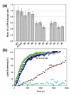
Functional characterization of mutant actins. a) Ratios of ε-ATP exchange rates of mutant actins to that in WT actin. The ε-ATP exchange assays were performed as described in Materials and Methods. The ratios of the calculated rates are presented with the error bars indicating the mean error. For WT actin, the rate of ε-ATP exchange was 0.0157 +/− 0.0018 sec−1. b) Actin (5.0 μM) polymerization by MgCl2 (3.0 mM) as monitored by light scattering at 325 nm. The plots were normalized to reveal kinetic differences in the polymerization reactions. For clarity, only representative polymerization data are given: for WT (overlapping with C41, C47) in black, for C49 (overlapping with C48, C45, and C42) in blue, for C46 in green, and C43 in gray. Mutant (C40) in cyan completed its polymerization at 6000 seconds under these conditions.
Figure 3
Figure 3
Disulfide cross-links between D-loop and C265 and C374. a) SDS-PAGE (non-reducing) of representative disulfide or MTS-6 cross-linked yeast F-actin co-polymers of D-loop mutants with C374 and C265 actins. The cross-linked pairs are indicated above the columns. The (−) and (+) signs indicate the presence or absence of 20 μMs of phalloidin, CuSO4, or MTS6 in the reaction mixtures. The time points at which the aliquots were removed are indicated in each panel. b) Stabilization of filaments by phalloidin increases the rates of longitudinal cross-linking. The observed rate constants in the presence (white circles) and absence (black circles) of phalloidin for the oxidation of co-polymers C43-C374 (on the right) and C50-C374 (on the left) were calculated by fitting the percentage of monomers to a first order exponential decay equation. The ratios of cross-linking rates in the presence of phalloidin to those in the absence of phalloidin were 1.44+/−0.12 and 1.41+/−0.14 for C43-C374 and C50-C374 filaments, respectively. c) The percentage of representative cross-linked dimers formed between D-loop and H-loop mutants in 30 seconds. The cross-linking products were analyzed on non-reducing SDS-PAGE after treatment with 2.0 mM NEM. The error bars represent the mean percentage error and the mean values are given inside the bars.
Figure 4
Figure 4
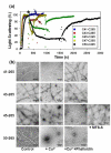
Effect of disulfide cross-links between D-loop and C374 on filament stability. a) Light scattering experiments detect the effect of disulfide cross-links between D-loop and C374 on F-actin. Co-polymers of D-loop mutants with C374 were polymerized with 3.0 mM MgCl2 and then 20 μM CuSO4 (indicated by star) was added to catalyze disulfide bond formation (observed on SDS-PAGE gels). Upon cross-linking completion, 10 mM TCEP (indicated by arrow) was added to the mixture to reduce the disulfide bonds. For easier comparison of the cross-linking effects, all light scattering data were normalized to 100%. b) Electron micrographs of yeast actin co-polymers before and after disulfide cross-linking. Micrographs of control (polymerized with 3 mM MgCl2) and disulfide cross-linked filaments are in the first and second columns; phalloidin induced recovery of the cross-linked filaments is shown in the third column. The rows are labeled with the corresponding co-polymer names. The MTS-6 cross-linked C45-C265 co-polymer micrograph is also shown. The scale bar corresponds to 0.1 micron in each image.
Figure 5
Figure 5
Effect of disulfide cross-links between D-loop and C265 on filament stability. a) Light scattering experiments detect the effect of disulfide cross-links between D-loop and C265 on F-actin. All experiments were performed as described in legend to Fig. 4a. The star and arrow denote the addition of 20 μM CuSO4 and 10 mM TCEP, respectively. b) Electron micrographs of yeast actin co-polymers before and after disulfide cross-linking. Micrographs of control and disulfide cross-linked filaments are in the first and second columns; phalloidin induced recovery of the cross-linked filaments is shown in the third column. The MTS-6 cross-linked C45-C265 co-polymer micrograph is also shown. The scale bar corresponds to 0.1 micron in each image.
Figure 6
Figure 6
SDS-PAGE (non-reducing) and the effect of disulfide cross-links between D-loop and C169 on actin filaments. a) SDS-PAGE of disulfide cross-linked for 30 seconds and 10 minutes F-actin (10 μM) co-polymers of D-loop mutants with the C169 mutant in the presence of 20 μM phalloidin. The co-polymers are identified above the columns. b) Light scattering experiments detect the effect of disulfide cross-links between the D-loop and C169 on F-actin. All experiments were performed as described in legend to Figure 4. The star and arrows denote the addition of 20 μM CuSO4 and 10 mM TCEP, respectively. The right column shows the EM images of oxidized F-actin copolymers of C41-C169 (c), C45-C169 (d), and C47-C169 (f). The EM image of filaments regenerated with the addition of phalloidin to aggregates shown in (d) is given in (e).
Figure 7
Figure 7
The minimum and mean Cα-Cα distances between the D-loop residues and C374 (a), C265 (b), and C169 (c) based on distances from the Cong actin scruin bundle model and molecular dynamics simulations of Holmes and Oda (2009) F-actin models. The C374 distance values are missing for Cong model since the C-terminus was not present in that model. The cross-links listed in Table 1 are color coded here according to their classification as having a minor (no change in filament morphology, green box), moderate (causing partial filament destruction, orange box) or severe phenotype (causing filament destruction, red box). “Mean” is the time averaged mean distance among the cross-linked pairs during simulations and “Min” is the minimum distance observed among the cross-linked residues during the simulations.

References

    1. Kabsch W, Mannherz HG, Suck D, Pai EF, Holmes KC. Atomic structure of the actin:DNase I complex. Nature. 1990;347:37–44. - PubMed
    1. Tirion MM, ben-Avraham D. Normal mode analysis of G-actin. J Mol Biol. 1993;230:186–95. - PubMed
    1. Graceffa P, Dominguez R. Crystal structure of monomeric actin in the ATP state. Structural basis of nucleotide-dependent actin dynamics. J Biol Chem. 2003;278:34172–80. - PubMed
    1. McLaughlin PJ, Gooch JT, Mannherz HG, Weeds AG. Structure of gelsolin segment 1-actin complex and the mechanism of filament severing. Nature. 1993;364:685–92. - PubMed
    1. Otterbein LR, Cosio C, Graceffa P, Dominguez R. Crystal structures of the vitamin D-binding protein and its complex with actin: structural basis of the actin-scavenger system. Proc Natl Acad Sci U S A. 2002;99:8003–8. - PMC - PubMed

Publication types

MeSH terms

LinkOut - more resources