. 2010 Jan 30;186(1):18-24.
doi: 10.1016/j.jneumeth.2009.10.017. Epub 2009 Nov 10.

High-speed X-ray video demonstrates significant skin movement errors with standard optical kinematics during rat locomotion

Affiliations

High-speed X-ray video demonstrates significant skin movement errors with standard optical kinematics during rat locomotion

Jay M Bauman et al. J Neurosci Methods. .

Abstract

The sophistication of current rodent injury and disease models outpaces that of the most commonly used behavioral assays. The first objective of this study was to measure rat locomotion using high-speed X-ray video to establish an accurate baseline for rat hindlimb kinematics. The second objective was to quantify the kinematics errors due to skin movement artefacts by simultaneously recording and comparing hindlimb kinematics derived from skin markers and from direct visualization of skeletal landmarks. Joint angle calculations from skin-derived kinematics yielded errors as high as 39 degrees in the knee and 31 degrees in the hip around paw contact with respect to the X-ray data. Triangulation of knee position from the ankle and hip skin markers provided closer, albeit still inaccurate, approximations of bone-derived, X-ray kinematics. We found that soft tissue movement errors are the result of multiple factors, the most impressive of which is overall limb posture. Treadmill speed had surprisingly little effect on kinematics errors. These findings illustrate the significance and context of skin movement error in rodent kinematics.


Figures

Figure 1
Figure 1
Sagittal plane kinematic model of rat hindlimb. Six tantalum markers (filled circles) were placed over bone landmarks corresponding to (proximal to distal): caudal ischium, greater trochanter, lateral epicondyle of the femur, lateral malleolus of the fibula, fourth metatarsal head, and the fourth distal phalanx. Included joint angles for hip, knee, ankle and metatarsophalangeal joints along with limb orientation were calculated.
Figure 2
Figure 2
Schematic of experimental set-up consisting of custom x-ray tube, rat on treadmill, image intensifier, high-speed digital camera, and computer interface.
Figure 3
Figure 3
Stick figures of the hindlimb showing average joint center positions calculated from bone-derived (gray), skin-derived (solid black) and triangulated (dashed) kinematics methods. Four specific times during the stride cycle are shown for four animals walking at 39.83 cm/s. Data are averaged over 44 gait cycles.
Figure 4
Figure 4
Mean kinematics for the hip (A), knee (B), and ankle (C) joints calculated from bone-derived (gray±1SD), skin-derived (solid) and triangulated (dashed) kinematics methods for the same representative animals as in Figure 3. Vertical line indicates end of stance phase and beginning of swing phase.
Figure 5
Figure 5
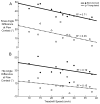
Knee (A) and hip (B) joint angle error at foot contact as a function of treadmill speed due to skin-derived (solid) and triangulated (dashed) kinematics methods. Joint angle error was defined as the difference with respect to bone-derived kinematics. Each symbol represents the average of all animals tested at that particular speed.
Figure 6
Figure 6
RMS error in knee joint position estimated from skin-derived (solid) and triangulated (dashed) methods as a function of limb orientation. Mean trajectories are pooled from all six rats across all treadmill speeds. Circles represent paw contact and the beginning of stance phase (thick lines) and end of the swing phase (thin lines). Arrows indicate flow of time over the stride cycle.
Figure 7
Figure 7
Mean resultant knee (A) and hip (B) joint acceleration trajectories calculated from both skin-derived (black) and bone-derived (gray) methods pooled for all rats across all speeds. Vertical line indicates transition from stance to swing.
Figure 8
Figure 8
Maximum knee (A) and hip (B) joint acceleration values calculated from both skin-derived (black) and bone-derived (gray) methods as a function of treadmill speed. Each symbol represents the average of all animals tested at that particular speed.
Figure 9
Figure 9
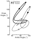
Mean interjoint coordination patterns of the knee joint angle as a function of ankle joint angle calculated from bone-derived (gray), skin-derived (solid black) and triangulated (dashed) kinematics methods for four animals walking at 39.8 cm/s. Circles represent paw contact. Arrows indicate flow of time over the stride cycle.

References

    1. Alexander EJ, Andriacchi TP. Correcting for deformation in skin-based marker systems. Journal of Biomechanics. 2001;34:355–61. - PubMed
    1. Anon . Washington, DC: U.S. Government Printing Office; 2001. Annual Report on Animal Welfare Act (AWA) Administration and Enforcement Activities In USDA.
    1. Bain JR, Mackinnon SE, Hunter DA. Functional evaluation of complete sciatic, peroneal, and posterior tibial nerve lesions in the rat. Plast Reconstr Surg. 1989;83:129–38. - PubMed
    1. Ballermann M, Tse AD, Misiaszek JE, Fouad K. Adaptations in the walking pattern of spinal cord injured rats. J Neurotrauma. 2006;23:897–907. - PubMed
    1. Banks SA, Fregly BJ, Boniforti F, Reinschmidt C, Romagnoli S. Comparing in vivo kinematics of unicondylar and bi-unicondylar knee replacements. Knee Surg Sports Traumatol Arthrosc. 2005;13:551–6. - PubMed

Publication types

MeSH terms

LinkOut - more resources