. 2010 Jan;38(1):46-54.
doi: 10.1016/j.exphem.2009.11.001.

Clonal analysis and hierarchy of human bone marrow mesenchymal stem and progenitor cells

Affiliations

Clonal analysis and hierarchy of human bone marrow mesenchymal stem and progenitor cells

C Chang I Lee et al. Exp Hematol. 2010 Jan.

Abstract

Objective: This study was performed to assess adult human bone marrow mesenchymal stem/progenitor cells at a single-cell level and to determine a hierarchy based on proliferative potential.

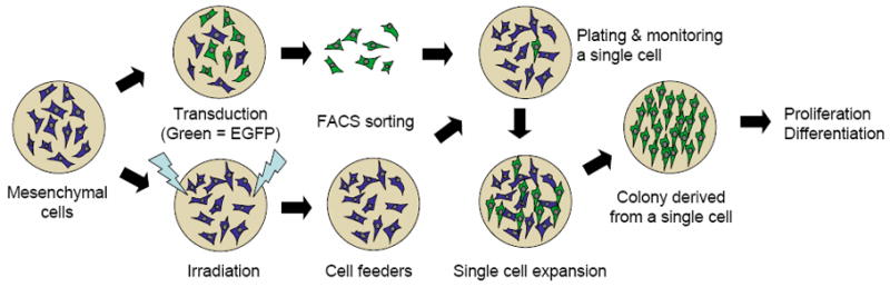
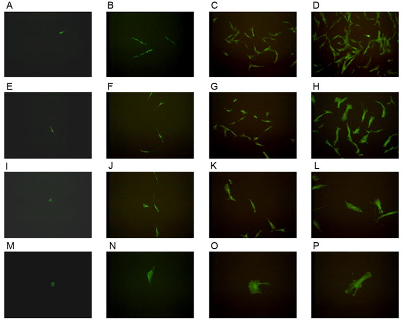
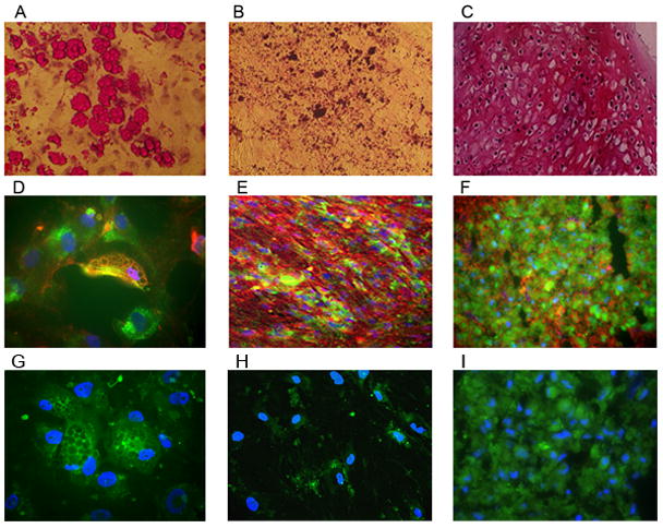
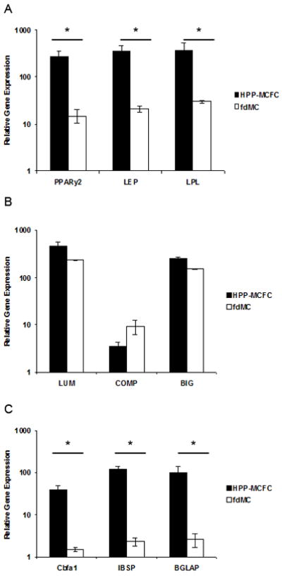
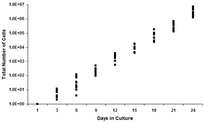
Materials and methods: Adult bone marrow mesenchymal cells expressing the enhanced green fluorescent protein (EGFP) were sorted as single cells into 24-well plates, each well confirmed with single EGFP-positive cells by fluorescence microscopy, and counted every 3 days. Colonies derived from single cells were expanded then sorted and evaluated using established differentiation protocols for adipogenic, chondrogenic, and osteogenic lineages. Cells were further analyzed by real-time reverse transcription polymerase chain reaction (RT-PCR) (peroxisome proliferator-activated receptor[PPAR]-gamma2, LEP, LPL, LUM, COMP, BIG, RUNX2, IBSP, BGLAP) and immunocytochemistry (PPAR-gamma1/2, collagen II, bone sialoprotein II) specific for trilineage differentiation.

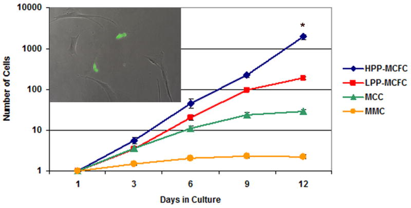
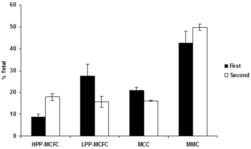
Results: Bone marrow mesenchymal cells were found to contain high proliferative potential (HPP) mesenchymal colony-forming cells (MCFC) (7%), low proliferative potential (LPP) MCFC (29%), mesenchymal cell clusters (MCC, 26%), and mature mesenchymal cells (MMC, 38%). All LPP-MCFC, MCC, and MMC colonies reached senescence at the end of the evaluation period. However, HPP-MCFC continued to grow, showed differentiation toward all three lineages, and demonstrated the capacity to give rise to secondary HPP-MCFC upon replating at a clonal level.

Conclusion: These findings suggest that there is a low frequency of bone marrow-derived HPP-MCFC that can both self-renew at a single-cell level and differentiate toward multiple lineages of mesenchymal origin.


Figures

Figure 1
Figure 1. Plating Protocol
The overall plan for cell plating is shown.
Figure 2
Figure 2. Fluorescence images of mesenchymal cell growth
Images of EGFP expressing cells obtained on day 1 (A, E, I, M) post-sorting, day 3 (B, F, J, N), day 6 (C, G, K, O), and day 9 (D, H, L, P). Four distinct cell populations derived from single mesenchymal cells included high proliferative potential-mesenchymal colony forming cells (HPP-MCFC; A–D), low proliferative potential-mesenchymal colony forming cells (LPP-MCFC; E–H), mesenchymal cell clusters (MCC; I–L), and mature mesenchymal cells (MMC; M–P) which were observed over time in culture. Images shown in each row were taken from the same well over time.
Figure 3
Figure 3. Growth of single mesenchymal cells
Single fluorescent mesenchymal cells expressing EGFP (green, insert) were sorted into 24-well plates with irradiated mesenchymal cells as feeders (EGFP-negative). The insert demonstrates the presence of two EGFP-expressing cells (day 3) within the irradiated feeder cells. EGFP-positive cells were counted every 3 days for 12 days. Wells containing ≥2 cells were excluded from the study. Single HPP-MCFC showed a significantly higher growth rate compared to LPP-MCFC, MCC, or MMC (p<0.05). All LPP-MCFC, MCC, and MMC reached senescence, whereas HPP-MCFC continued to proliferate. *p<0.05.
Figure 4
Figure 4. Differentiation of mesenchymal cell clonal colonies
Colonies of fluorescent HPP-MCFC were differentiated toward adipogenic (A, D), osteogenic (B, E), and chondrogenic (C, F) lineages. Adipogenic-differentiated cells were stained with Oil Red-O (red channel only, A, 10X objective) and co-expressed PPAR γ1/γ2 (red, D, 40X objective) with the EGFP-HPP-MCFC. Osteogenic-differentiated cells were stained with Von Kossa (red channel only, B, 10X objective) and co-expressed BSP (red, E, 20X objective) with the EGFP-HPP-MCFC. Sections of chondrogenic-differentiated pellets were stained with Safranin-O (red channel only, C, 10X objective) and co-expressed Collagen II (red, F, 20X objective) with the EGFP-HPP-MCFC. No staining (other than the EGFP of the HPP-MCFC progeny) was observed in secondary antibody controls (G–I). Blue = DAPI staining of nuclei.
Figure 5
Figure 5. PCR analysis of differentiated HPP-MCFC
Genes specific for adipogenic (A), chondrogenic (B), and osteogenic (C) lineages were analyzed after sorting EGFP-positive differentiated HPP-MCFC from feeder mesenchymal cells (fdMC, EGFP-negative). Significantly greater levels of adipogenic (A) and osteogenic (C) gene expression were observed in HPP-MCFC compared to fdMC (p<0.05). This effect was not as profound for chondrogenic (B) differentiation when compared to the other two lineages. *p<0.05.
Figure 6
Figure 6. Growth of single mesenchymal cells after sorting
HPP-MCFC from the initial sorting of single cells were pooled, sorted, and monitored for 12 days. The percentage of HPP-MCFC (>9 population doublings) and MMC (≤3 population doublings) appeared to increase from the first sort to the second sort, while LPP-MCFC (>6–9 population doublings) and MCC (>3–6 population doublings) were observed to decrease.
Figure 7
Figure 7. Cumulative growth of HPP-MCFC
Single HPP-MCFC proliferated to 3.8×10⁶ ± 6.6×10⁵ cells over 24 days.

References

    1. McNiece IK, Bertoncello I, Kriegler AB, Quesenberry PJ. Colony-forming cells with high proliferative potential (HPP-CFC) Int J Cell Cloning. 1990;8:146–160. - PubMed
    1. Yoder MC, Du XX, Williams DA. High proliferative potential colony-forming cell heterogeneity identified using counterflow centrifugal elutriation. Blood. 1993;82:385–391. - PubMed
    1. Bradley TR, Hodgson GS. Detection of primitive macrophage progenitor cells in mouse bone marrow. Blood. 1979;54:1446–1450. - PubMed
    1. Srour EF, Brandt JE, Briddell RA, Grigsby S, Leemhuis T, Hoffman R. Long-term generation and expansion of human primitive hematopoietic progenitor cells in vitro. Blood. 1993;81:661–669. - PubMed
    1. Ingram DA, Mead LE, Tanaka H, et al. Identification of a novel hierarchy of endothelial progenitor cells using human peripheral and umbilical cord blood. Blood. 2004;104:2752–2760. - PubMed

Publication types