. 2010 Jan;36(1):145-57.
doi: 10.1016/j.ultrasmedbio.2009.08.009.

Ultrasound-triggered release of recombinant tissue-type plasminogen activator from echogenic liposomes

Affiliations

Ultrasound-triggered release of recombinant tissue-type plasminogen activator from echogenic liposomes

Denise A B Smith et al. Ultrasound Med Biol. 2010 Jan.

Abstract

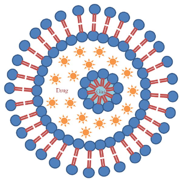
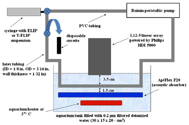
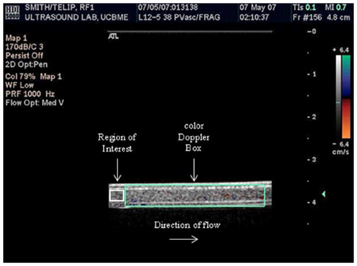
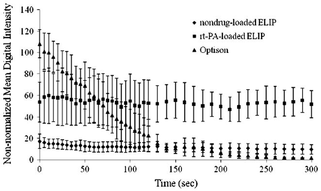
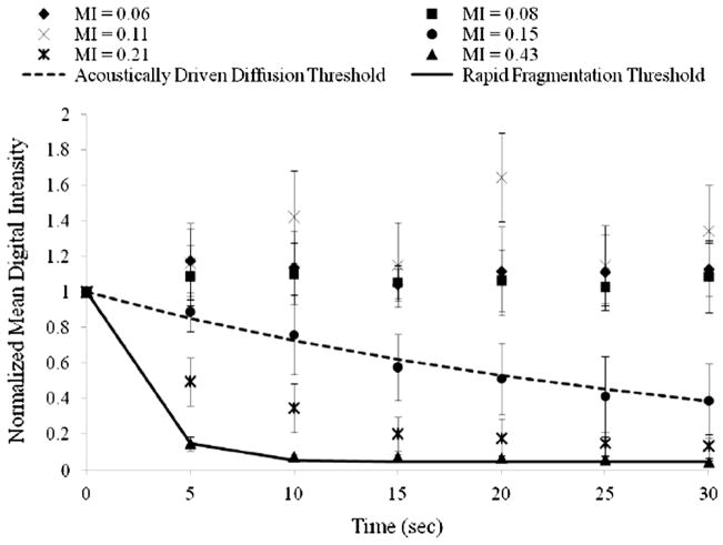
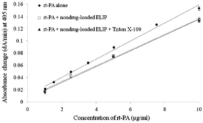
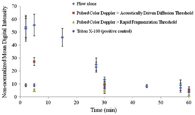
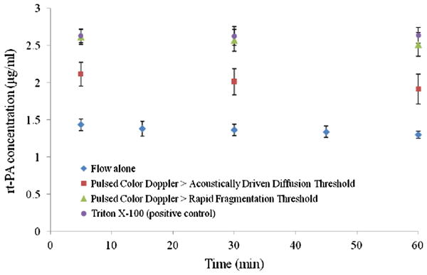
Echogenic liposomes (ELIP) were developed as ultrasound-triggered targeted drug or gene delivery vehicles (Lanza et al. 1997; Huang et al. 2001). Recombinant tissue-type plasminogen activator (rt-PA), a thrombolytic, has been loaded into ELIP (Tiukinhoy-Laing et al. 2007). These vesicles have the potential to be used for ultrasound-enhanced thrombolysis in the treatment of acute ischemic stroke, myocardial infarction, deep vein thrombosis or pulmonary embolus. A clinical diagnostic ultrasound scanner (Philips HDI 5000; Philips Medical Systems, Bothell, WA, USA) equipped with a linear array transducer (L12-5) was employed for in vitro studies using rt-PA-loaded ELIP (T-ELIP). The goal of this study was to quantify ultrasound-triggered drug release from rt-PA-loaded echogenic liposomes. T-ELIP samples were exposed to 6.9-MHz B-mode pulses at a low pressure amplitude (600 kPa) to track the echogenicity over time under four experimental conditions: (1) flow alone to monitor gas diffusion from the T-ELIP, (2) pulsed 6.0-MHz color Doppler exposure above the acoustically driven threshold (0.8 MPa) to force gas out of the liposome gently, (3) pulsed 6.0-MHz color Doppler above the rapid fragmentation threshold (2.6 MPa) or (4) Triton X-100 to rupture the T-ELIP chemically as a positive control. Release of rt-PA for each ultrasound exposure protocol was assayed spectrophotometrically. T-ELIP were echogenic in the flow model (5 mL/min) for 30 min. The thrombolytic drug remained associated with the liposome when exposed to low-amplitude B-mode pulses over 60 min and was released when exposed to color Doppler pulses or Triton X-100. The rt-PA released from the liposomes had similar enzymatic activity as the free drug. These T-ELIP are robust and echogenic during continuous fundamental 6.9-MHz B-mode imaging at a low exposure output level (600 kPa). Furthermore, a therapeutic concentration of rt-PA can be released by fragmenting the T-ELIP with pulsed 6.0-MHz color Doppler ultrasound above the rapid fragmentation threshold (1.59 MPa). (E-mail: denise.smith@uc.edu).


Figures

Figure 1
Figure 1
Proposed structure of an echogenic liposome (ELIP) with an outer phospholipid bilayer. A lipid monolayer shell surrounds a gas bubble in the internal aqueous compartment of the liposome. The gas core provides the acoustic impedance mismatch necessary for ELIP to be used as contrast agents in diagnostic ultrasound imaging. Note that the aqueous contents, shown as an encapsulated drug, may be triggered to release by an ultrasound pulse.
Figure 2
Figure 2
Schematic of the in vitro flow model. ELIP or T-ELIP suspensions were pumped through tubing using a peristaltic pump at 5 ml/min. A 10 cm portion of the latex tubing was submersed in an aquarium tank of 0.2 μm filtered, deionized water and the transducer face of a Philips L12-5 linear array was immersed 3.5 cm above the model. Acoustic absorber material was placed 1.5 cm below the model to prevent the reflection of ultrasound. Aquarium heaters were placed inside the tank to keep water temperature at 37 °C. T-ELIP suspensions were collected into a disposable cuvette at various timed points using a three-way valve.
Figure 3
Figure 3
Freeze-captured image of T-ELIP circulating in the flow model. The probe was transmitting maximal color Doppler pulses (fc = 6.0 MHz) with simultaneous fundamental B-mode pulses (fc = 6.9 MHz). The fixed color Doppler window (large teal box) encompassed the entire lumen of the latex tube and a 12-mm2 region of interest was drawn within the lumen of the latex tube (small white box). Note that flow was from left to right in this image. fc = center frequency
Figure 4
Figure 4
Diameter size distribution of T-ELIP assessed by a Beckman-Coulter Multisizer 3 counter using a 20-μm aperture. Note the peak number density (count/ml) of T-ELIP is at a diameter of 1.6 μm.
Figure 5
Figure 5
Loss of echogenicity of three different contrast agent suspensions during scanned harmonic B-mode imaging (fc = 4.5 MHz) at MI = 0.04 (Pr = 0.12 MPa) shown as non-normalized mean digital intensity over time. MI = Mechanical Index, Pr = peak rarefactional pressure, fc = center frequency
Figure 6
Figure 6
Graph for 6.0-MHz duplex Doppler imaging (PD = 3.33 μs, PRF = 1.25 kHz) of normalized digital intensity as a function of time for prolonged exposure to peak rarefactional pressures (Pr) of (◆) 0.22 MPa, (■) 0.30 MPa, (×) 0.41 MPa, (●) 0.56 MPa, (*) 0.78 MPa, and (▲) 1.59 MPa of rt-PA-loaded echogenic liposome suspensions (N = 5). Note that the acoustically driven diffusion threshold was defined at Pr = 0.56 MPa (MI = 0.15) and the rapid fragmentation threshold was defined at Pr = 1.59 MPa (MI = 0.43). The smoothed lines represent the least squares fit of the data to an exponential decay function of the form Aekt + N. PD = pulse duration, PRF = pulse repetition frequency, MI = Mechanical Index
Figure 7
Figure 7
Nondrug-loaded rt-PA concentration curves for the absorbance change (ΔA/min) at 405 nm versus rt-PA alone (y = 0.0148x + 0.0108, R2 = 0.99), rt-PA plus nondrug-loaded ELIP (y = 0.0128x + 0.0085, R2 = 1.00), or rt-PA plus nondrug-loaded ELIP and Triton X-100 (y = 0.0129x + 0.0066, R2 = 1.00). Note that the rt-PA concentrations chosen were from 1 to 10 μg/ml or the therapeutic concentration range in humans.
Figure 8
Figure 8
Loss of echogenicity over time for T-ELIP suspensions at 37 °C and 5 ml/min exposed to continuously scanned fundamental B-mode pulses at a center frequency of 6.9-MHz (MI = 0.04, Pr = 600 kPa) in four separate exposures conditions; 1) flow alone, 2) color 6.0-MHz Doppler pulses above acoustically driven threshold (MI = 0.22, Pr = 0.8 MPa), 3) color 6.0-MHz Doppler pulses above rapid fragmentation threshold (MI = 0.7, Pr = 2.6 MPa), and 4) Triton X-100 (0.07% v/v). The T-ELIP were reconstituted at time zero. Note that the data shown at time points (2, 37 and 57 min) correspond to the echogenicity of T-ELIP suspensions (N = 5) with flow alone before being exposed to the color Doppler pulses for the next three minutes. Pr = peak rarefactional pressure, MI = Mechanical Index
Figure 9
Figure 9
Concentration of rt-PA over time for T-ELIP suspensions at 37 °C and 5 ml/min exposed to continuously scanned fundamental B-mode pulses at a center frequency of 6.9-MHz (MI = 0.04, Pr = 600 kPa) in four separate exposures conditions; 1) flow alone, 2) color 6.0-MHz Doppler pulses above acoustically driven threshold (MI = 0.22, Pr = 0.8 MPa), 3) color 6.0-MHz Doppler pulses above rapid fragmentation threshold (MI = 0.7, Pr = 2.6 MPa), and 4) Triton X-100 (0.07% v/v). The T-ELIP were reconstituted at time, 0. Pr = peak rarefactional pressure, MI = Mechanical Index

References

    1. Alexandrov AV. Ultrasound enhanced thrombolysis for stroke. Seminars in Cerebrovascular Diseases and Stroke. 2006;5:106–110.
    1. Apfel RE, Holland CK. Gauging the likelihood of cavitation from short-pulse, low-duty cycle diagnostic ultrasound. Ultrasound Med Biol. 1991;17:179–185. - PubMed
    1. Bevan DP, Karshafian R, Burns PN. The influence of fragmentation on the acoustic response from shrinking bubbles. Ultrasound Med Biol. 2008;34 (7):1152–1162. - PubMed
    1. Borden MA, Kruse DE, Caskey CF, Zhao S, Dayton PA, Ferrara KW. Influence of lipid shell physiochemical properties on ultrasound-induced microbubble destruction. IEEE Trans Ultrason Ferroelectr Freq Control. 2005;52:1992–2002. - PMC - PubMed
    1. Bouakaz A, de Jong N. WFUMB safety symposium on echo-contrast agents: Nature and types of ultrasound contrast agents. Ultrasound Med Biol. 2007;33:187–196. - PubMed

Publication types