. 2009 Oct 30;326(5953):734-6.
doi: 10.1126/science.1178258.

Hemagglutinin receptor binding avidity drives influenza A virus antigenic drift

Affiliations

Hemagglutinin receptor binding avidity drives influenza A virus antigenic drift

Scott E Hensley et al. Science. .

Abstract

Rapid antigenic evolution in the influenza A virus hemagglutinin precludes effective vaccination with existing vaccines. To understand this phenomenon, we passaged virus in mice immunized with influenza vaccine. Neutralizing antibodies selected mutants with single-amino acid hemagglutinin substitutions that increased virus binding to cell surface glycan receptors. Passaging these high-avidity binding mutants in naïve mice, but not immune mice, selected for additional hemagglutinin substitutions that decreased cellular receptor binding avidity. Analyzing a panel of monoclonal antibody hemagglutinin escape mutants revealed a positive correlation between receptor binding avidity and escape from polyclonal antibodies. We propose that in response to variation in neutralizing antibody pressure between individuals, influenza A virus evolves by adjusting receptor binding avidity via amino acid substitutions throughout the hemagglutinin globular domain, many of which simultaneously alter antigenicity.


Figures
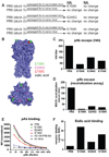
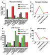
Fig. 1
Fig. 1. In vivo influenza virus passaging selects for mutants with altered binding avidity
(A) HA and NA genes were sequenced in lung homogenates from 3 independent PR8 stocks serially passaged in vaccinated and naïve Swiss mice. (B) Location of in vivo selected HA amino acid substitutions in mutant viruses. (C) PR8 and mutant viruses were tested for escape from anti-PR8 polyclonal antibodies by HAI using turkey erythrocytes or (D) virus neutralization assays using MDCK cells. Data are expressed as inverse dilutions of serum and are representative of three (HAI) or two (virus neutralization) experiments. Means +/− SEM are shown in panel D. (E) Polyclonal antibody binding to HA was assessed by flow cytometry after adding different dilutions of polyclonal antibody to L929 cells infected with the indicated virus followed by the addition of anti-mouse FITC. Shown is mean florescence intensity (MFI) after normalizing HA expression based on the binding of a mixture of Ca monoclonal antibodies or a NA specific monoclonal antibody (for the H3 HA/PR8 NA virus). Polyclonal antibodies bind nearly exclusively to HA, as inferred from their low binding to the H3 HA/PR8 NA infected cells. Data are representative of three independent experiments. (F) Cellular receptor binding avidities were determined by hemagglutination of turkey erythrocytes pre-treated with RDE. Data are expressed as the maximal amount of RDE that allowed full agglutination. Data are representative of three independent experiments.
Fig. 2
Fig. 2. Numerous HA amino acid substitutions simultaneously modulate receptor binding and escape from polyclonal antibodies
(A) Cellular avidities of 40 viruses with single HA amino acid substitutions were determined using RDE-treated turkey erythrocytes. The percentages of viruses within each antigenic group with altered binding are shown. (B) Individual mutants were tested for their abilities to escape PR8 polyclonal antibodies by HAI. Data are plotted as ability to escape polyclonal antibodies vs cellular receptor binding avidity. Means are represented as dots and the 95% confidence interval is represented by dashed lines. (C) Locations of HA amino acid substitutions that promote increased avidity are shown in yellow. (D) Net charge change of mutant viruses was determined. n = number of viruses in each group. Means +/− SEM are shown.
Fig. 3
Fig. 3. Removal of polyclonal antibody selection promotes decreased receptor binding of in vivo selected mutants
(A) Mutant viruses were tested for agglutination of RDE-treated turkey erythrocytes. REV=virus with amino acid reversion of original mutation. (B) Mutant viruses were tested for escape from PR8 polyclonal antibodies by HAI. Receptor binding avidities (C) and ability to escape PR8 polyclonal antibodies (D) were also determined for the virus that was isolated after further passage in heavily vaccinated mice.

Similar articles

Cited by

References

    1. Air GM, Laver WG, Webster RG. Contrib Microbiol Immunol. 1987;8:20. - PubMed
    1. Dimmock NJ. J Gen Virol. 1984;65:1015. - PubMed
    1. Knossow M, Skehel JJ. Immunol. 2006;119:1. - PMC - PubMed
    1. Caton AJ, Brownlee GG, Yewdell JW, Gerhard W. Cell. 1982;31:417. - PubMed
    1. Yewdell JW, Webster RG, Gerhard WU. Nature. 1979;279:246. - PubMed

Publication types

MeSH terms