. 2010 Jan 1;588(Pt 1):225-42.
doi: 10.1113/jphysiol.2009.181800. Epub 2009 Nov 9.

Increased Ca(2+) leak and spatiotemporal coherence of Ca(2+) release in cardiomyocytes during beta-adrenergic stimulation

Affiliations

Increased Ca(2+) leak and spatiotemporal coherence of Ca(2+) release in cardiomyocytes during beta-adrenergic stimulation

Jakob Ogrodnik et al. J Physiol. .

Abstract

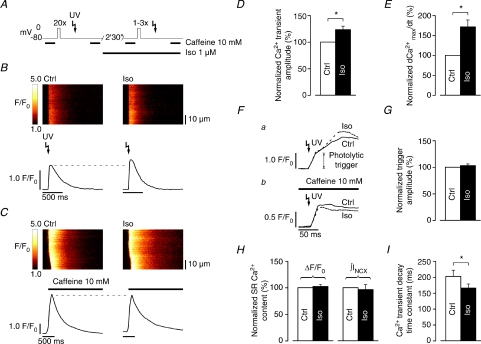
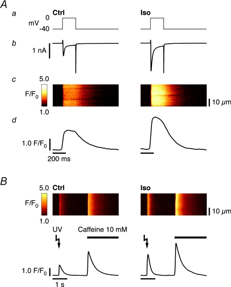
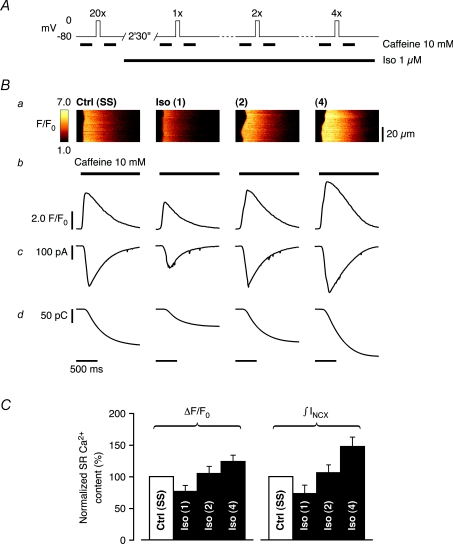
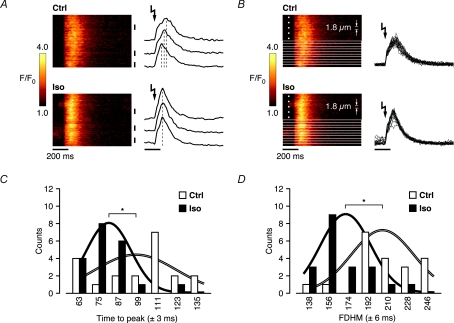
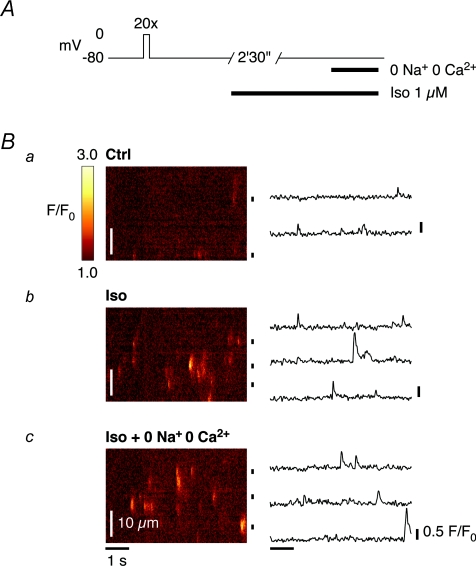
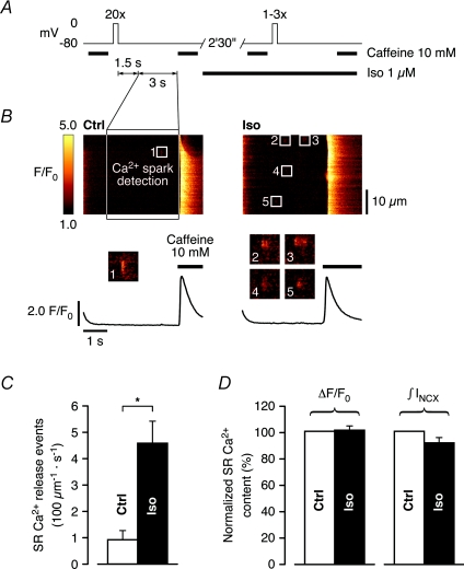
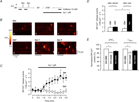
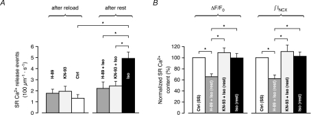
beta-Adrenergic receptor (beta-AR) stimulation of cardiac muscle has been proposed to enhance Ca(2+) release from the sarcoplasmic reticulum (SR) through ryanodine receptors (RyRs). However, the anticipated increase in RyR Ca(2+) sensitivity has proven difficult to study in intact cardiomyocytes, due to accompanying alterations in SR Ca(2+) content, inward Ca(2+) current (I(Ca)) and diastolic cytosolic Ca(2+) concentration ([Ca(2+)](i)). Here, we studied whole-cell Ca(2+) release and spontaneous Ca(2+) leak (Ca(2+) sparks) in guinea-pig ventricular myocytes with confocal Ca(2+) imaging before and during beta-AR stimulation by isoproterenol (Iso), but under otherwise nearly identical experimental conditions. The extent of SR Ca(2+) loading was controlled under whole-cell voltage-clamp conditions. UV flash-induced uncaging of Ca(2+) from DM-nitrophen was employed as an invariant trigger for whole-cell Ca(2+) release. At matched SR Ca(2+) content, we found that Iso enhanced the spatiotemporal coherence of whole-cell Ca(2+) release, evident from spatially intercorrelated release and accelerated release kinetics that resulted in moderately (20%) increased release amplitude. This may arise from higher RyR Ca(2+) sensitivity, and was also reflected in spontaneous SR Ca(2+) leak. At comparable SR Ca(2+) content and cytosolic [Ca(2+)](i), we observed an approximately 4-fold increase in Ca(2+) spark frequency in Iso that also appeared in quiescent cells within 2 min without increased SR Ca(2+) content. This was likely to have been mediated by Ca(2+)/calmodulin-dependent protein kinase (CaMKII), rather than cAMP dependent protein kinase (PKA). We conclude that Iso increases the propensity of RyRs to open, both in response to rapid elevations of [Ca(2+)](i) and at diastolic [Ca(2+)](i). While this could be beneficial in enhancing and synchronizing systolic whole-cell SR Ca(2+) release, the same behaviour could also be proarrhythmogenic during diastole.


Figures

Figure 3
Figure 3. UV flash-induced SR Ca2+ release is increased in Iso at matched SR Ca2+ content
A, whole-cell SR Ca2+ release induced by UV flash-photolysis of DM-nitrophen in control (SR loaded to steady-state), and again following an adapted preloading protocol (1–3 preloading steps) in Iso. Rapid application of 10 mm caffeine 2 s after the UV flash in both control and Iso was used to verify comparable SR Ca2+ content. B, whole-cell SR Ca2+ release was higher in Iso (right) compared to control (left), despite similar SR Ca2+ content (C). On average, whole-cell SR Ca2+ release was significantly stimulated to ΔF/F0: 123.4 ± 6.3% of control (n= 8 cells, D). E, Iso accelerated whole-cell SR Ca2+ release kinetics (d[Ca2+]max dt−1: 171.6 ± 17.0% of control, n= 8 cells). F, the amplitude of the initial (rapid) phase of UV flash-induced Ca2+ transients (a) confirmed that the photolytical trigger for whole-cell SR Ca2+ release was comparable in control and Iso, as did UV flash-induced Ca2+ transients recorded in the constant presence of 10 mm caffeine (b, data from another cell). G, average amplitude of the photolytical trigger extracted from the biphasic upstroke of UV flash-induced Ca2+ transients (ΔF/F0: 102.9 ± 2.9% of control, n= 4 cells). H, SR Ca2+ content was reliably matched in these experiments (caffeine-induced ΔF/F0: 102.1 ± 3.6% and ∫INCX: 96.4 ± 9.1% of control, n= 8 cells). I, Iso also accelerated whole-cell Ca2+ transient decay kinetics (τ: 202.4 ± 19.0 ms in control to 165.8 ± 12.4 ms in Iso, n= 8 cells) (*P < 0.01).
Figure 1
Figure 1. Iso increases Ca2+ transients by enhancing ICa and elevating SR Ca2+ content
A, depolarizing step (200 ms, from −40 mV to 0 mV, a), recorded current (b), acquired line-scan image (c) and average temporal Ca2+ transient profile (d). Compared to control (left), 2 min 30 s of β-AR stimulation with 1 μm Iso activates larger ICa and increases CICR, resulting in a larger cytosolic Ca2+ transient (right). B, whole-cell SR Ca2+ release in response to UV flash-induced Ca2+ uncaging from DM-nitrophen, following identical preconditioning in control (left) and Iso (right), recorded in a different cell. Stimulation of SERCA activity in Iso leads to more SR Ca2+ loading (confirmed with rapid application of 10 mm caffeine), and a larger cytosolic UV flash-induced Ca2+ transient, and may mask direct functional modulation of the RyR resulting from β-AR stimulation.
Figure 2
Figure 2. SR Ca2+ content can be matched in Iso by adaptation of the preconditioning protocol
A, adjusting SR Ca2+ loading under our experimental conditions. Following initial emptying with caffeine, SR Ca2+ content was assessed in control after reloading to steady-state (SS) with 20 depolarizing steps (200 ms, from −80 mV to 0 mV, 0.5 Hz). In Iso, the SR was successively emptied and its content reassessed with caffeine after reloading with 1, 2 and 4 depolarizing steps. B, acquired line-scan images of caffeine-induced Ca2+ release after the number of depolarizing steps indicated in A (a), corresponding Ca2+ transient profiles (b), accompanying INCX (c) and their integrals (d). As expected, SR Ca2+ content increased with the number of preloading steps in Iso. C, relative SR loading conditions normalized to control, as estimated from the amplitude of the Ca2+ transient (ΔF/F0) and integral of the INCX (∫INCX) accompanying rapid caffeine exposure after 1, 2 and 4 loading steps. On average, SR reloading with 2 depolarizing steps in Iso best matched SR Ca2+ content in steady-state in control (ΔF/F0: 104.8 ± 11.0% and ∫INCX: 106.0 ± 12.1% of control, n= 5 cells), although in some cells 1 or even 4 depolarizing steps yielded a closer match.
Figure 4
Figure 4. Increased spatial synchronization of whole-cell SR Ca2+ release in Iso is revealed at near-threshold triggers
A, near-threshold SR Ca2+ release kinetics exhibited clear spatial inhomogeneities in control, as indicated by the temporal Ca2+ transient profiles from 3 different (subcellular) regions. Iso synchronized SR Ca2+ release throughout the cell, as reflected by the coordinated peaks of the corresponding subcellular Ca2+ transient profiles. B, the line-scan images in control and Iso in A were divided into 20 equal parts (each 1.8 μm wide), on which a detailed analysis of the temporal characteristics was performed. C, the distribution of the time to peak in the subcellular regions revealed a significantly shorter average time to peak in Iso (99.3 ± 5.4 ms in control to 78.9 ± 3.0 ms in Iso), and was less spread in time throughout the cell (reflected by the relative widths and amplitudes of the representative, integral-normalized Gaussians). D, accelerated release and decay kinetics in Iso are also reflected in shorter average full duration at half-maximum (FDHM) amplitude throughout the cell (207.6 ± 6.3 ms in control to 168.0 ± 5.0 ms in Iso) (*P < 0.01).
Figure 5
Figure 5. Ca2+ sparks are more frequent in quiescent guinea-pig ventricular myocytes in Iso
A, the occurrence of Ca2+ sparks in voltage-clamped ventricular myocytes was studied in quiescent cells, after loading of the SR with Ca2+ to steady-state in control conditions. B, Ca2+ sparks were relatively rare in control (a), but the number of readily visible Ca2+ sparks markedly increased during β-AR stimulation with 1 μm Iso for 2 min 30 s (b). Their appearance persisted when the extracellular solution was exchanged for a solution without Na+ or Ca2+ (0 Na+, 0 Ca2+) (c), indicating that the SR Ca2+ leak is spontaneous and independent of extracellular Ca2+ (i.e. not triggered by any Ca2+ influx).
Figure 6
Figure 6. Spontaneous SR Ca2+ release is increased at matched SR Ca2+ content and comparable diastolic [Ca2+]i in Iso
A, spontaneous SR Ca2+ release was compared in control and in Iso at matched SR Ca2+ content (cf. experimental protocol described in Fig. 3). Ca2+ sparks were recorded in control (SR loaded to steady-state), and again following an adapted preloading protocol (1–3 preloading steps) in Iso. Analysis was performed on Ca2+ sparks during a 3 s long period starting 1.5 s after the final repolarization to −80 mV. Subsequent rapid application of 10 mm caffeine was used to verify comparable SR Ca2+ content. B, Ca2+ sparks after repolarization and complete decay of [Ca2+]i to diastolic levels were generally more frequent in Iso (a single spark in control, left, to 4 in Iso, right). C, on average, Ca2+ spark frequency increased ∼5-fold in Iso (0.92 ± 0.34 s−1 (100 μm)−1 in control to 4.58 ± 0.83 s−1 (100 μm)−1 in Iso), in recordings where the SR Ca2+ content was matched to D, SR Ca2+ content was matched in these recordings (caffeine-induced ΔF/F0: 100.8 ± 3.0% and ∫INCX: 91.3 ± 3.8% of control) (n= 9 cells, *P < 0.01).
Figure 7
Figure 7. Ca2+ spark frequency increases rapidly in quiescent cells in Iso without significantly altering SR Ca2+ content
A, experimental protocol to study the time course of spontaneous SR Ca2+ release and how it affects SR Ca2+ content during rest. SR Ca2+ content was assessed with caffeine after loading of the SR with Ca2+ to steady-state in control. Following reloading of the SR to steady-state, cells were left to rest for 3 min (30 s in control + 2 min 30 s in Iso, or 3 min in control), after which SR Ca2+ content was again assessed with caffeine. B and C, a progressive increase in Ca2+ spark frequency appeared after ∼30 s superfusion with Iso, reaching an ∼4-fold higher level within 2 min (a, b′, c′). Cells in control did not exhibit any obvious change in Ca2+ spark frequency during rest (a, b, c). D, Ca2+ spark frequency was not significantly lower after rest in control (0.93 ± 0.21 s−1 (100 μm)−1 to 0.66 ± 0.19 s−1 (100 μm)−1). After rest in Iso, Ca2+ spark frequency increased from 1.05 ± 0.13 s−1 (100 μm)−1 to 4.08 ± 0.47 s−1 (100 μm)−1; data were pooled from the initial 3 points and the final 3 points from both groups). E, SR Ca2+ content decreased significantly after rest in control (caffeine-induced ΔF/F0: 82.0 ± 3.3% and ∫INCX: 81.4 ± 4.7% of initial SR Ca2+ content), whereas this loss of Ca2+ was more limited after rest in Iso (caffeine-induced ΔF/F0: 89.5 ± 3.5% and ∫INCX: 91.4 ± 5.5% of initial SR Ca2+ content) (control: n= 10 cells, Iso: n= 9 cells, *P < 0.01).
Figure 8
Figure 8. CaMKII rather than PKA mediates the increased spontaneous SR Ca2+ release in Iso
The time course of spontaneous SR Ca2+ release during rest was monitored following incubation with either the PKA inhibitor H-89 (5 μm) or the CaMKII inhibitor KN-93 (5 μm). H-89 or KN-93 was added to the extracellular solution immediately after reloading of the SR to steady-state. A, compared to Iso, which increased Ca2+ spark frequency from 1.32 ± 0.32 s−1 (100 μm)−1 to 4.92 ± 0.56 s−1 (100 μm)−1, treatment with either inhibitor almost completely suppressed the increase in Ca2+ spark frequency (from 1.77 ± 0.36 s−1 (100 μm)−1 in H-89 to 2.21 ± 0.57 s−1 (100 μm)−1 in H-89 + Iso, n.s., and from 1.95 ± 0.44 s−1 (100 μm)−1 in KN-93 to 2.44 ± 0.40 s−1 (100 μm)−1 in KN-93 + Iso, n.s.). B, during rest in KN-93 + Iso, SR Ca2+ content exhibited a tendency to increase in parallel, although this increase was not significant (caffeine-induced ΔF/F0: 108.8 ± 8.6% and ∫INCX: 110.9 ± 11.9% of initial SR Ca2+ content). During rest in H-89 + Iso, however, the loss of Ca2+ from the SR was dramatic (caffeine-induced ΔF/F0: 65.4 ± 4.9% and ∫INCX: 62.0 ± 6.5% of initial SR Ca2+ content). SR Ca2+ content was again unaffected after rest in Iso (caffeine-induced ΔF/F0: 99.6 ± 7.8% and ∫INCX: 102.7 ± 7.4% of initial SR Ca2+ content) (H-89 ± Iso: n= 8 cells, KN-93 ± Iso: n= 7 cells, Iso: n= 5 cells, *P < 0.01).

Comment in

Similar articles

Cited by

References

    1. Ai X, Curran JW, Shannon TR, Bers DM, Pogwizd SM. Ca2+/calmodulin-dependent protein kinase modulates cardiac ryanodine receptor phosphorylation and sarcoplasmic reticulum Ca2+ leak in heart failure. Circ Res. 2005;97:1314–1322. - PubMed
    1. Altamirano J, Bers DM. Voltage dependence of cardiac excitation-contraction coupling: unitary Ca2+ current amplitude and open channel probability. Circ Res. 2007;101:590–597. - PubMed
    1. Barrett SD. Software for scanning microscopy. Proc Roy Microsc Soc. 2002;37:164–174.
    1. Benkusky NA, Weber CS, Scherman JA, Farrell EF, Hacker TA, John MC, Powers PA, Valdivia HH. Intact β-adrenergic response and unmodified progression toward heart failure in mice with genetic ablation of a major protein kinase A phosphorylation site in the cardiac ryanodine receptor. Circ Res. 2007;101:819–829. - PubMed
    1. Berlin JR, Cannell MB, Lederer WJ. Cellular origins of the transient inward current in cardiac myocytes. Role of fluctuations and waves of elevated intracellular calcium. Circ Res. 1989;65:115–126. - PubMed

Publication types

MeSH terms