. 2010 Jan 1;588(Pt 1):125-37.
doi: 10.1113/jphysiol.2009.181107. Epub 2009 Nov 9.

Excitability parameters and sensitivity to anemone toxin ATX-II in rat small diameter primary sensory neurones discriminated by Griffonia simplicifolia isolectin IB4

Affiliations

Excitability parameters and sensitivity to anemone toxin ATX-II in rat small diameter primary sensory neurones discriminated by Griffonia simplicifolia isolectin IB4

Alistair Snape et al. J Physiol. .

Abstract

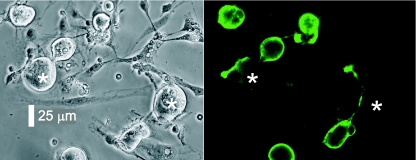
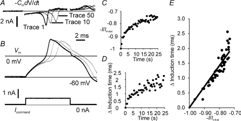
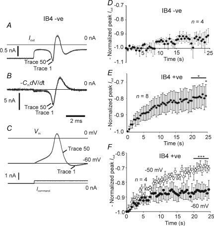
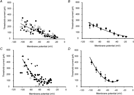
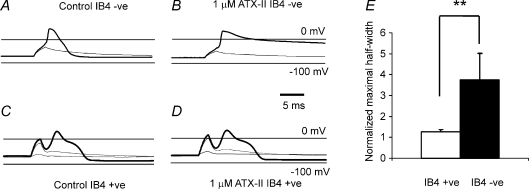
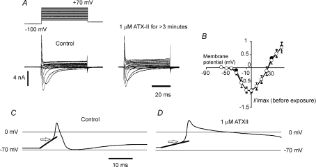
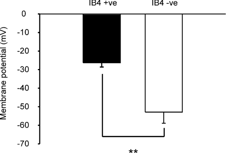
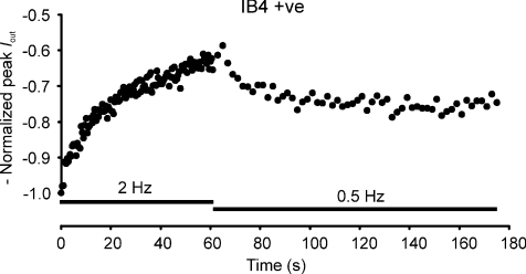
Sensory neurone subtypes (< or = 25 microm apparent diameter) express a variety of Na(+) channels, where expression is linked to action potential duration, and associated with differential IB4-lectin binding. We hypothesized that sensitivity to ATX-II might also discriminate neurones and report that 1 microm has negligible or small effects on action potentials in IB4 +ve, but dramatically increased action potential duration in IB4 ve, neurones. The toxin did not act on tetrodotoxin-resistant (TTX-r) Na(V)1.8 currents; discrimination was based on tetrodotoxin-sensitive (TTX-s) Na(+) channel expression. We also explored the effects of varying the holding potential on current threshold, and the effect of repetitive activation on action currents in IB4 +ve and ve neurones. IB4 +ve neurones became more excitable with depolarization over the range 100 to 20 mV, but IB4 ve neurones exhibited peak excitability near 55 mV, and were inexcitable at 20 mV. Eliciting action potentials at 2 Hz, we found that peak inward action current in IB4 +ve neurones was reduced, whereas changes in the current amplitude were negligible in most IB4 ve neurones. Our findings are consistent with relatively toxin-insensitive channels including Na(V)1.7 being expressed in IB4 +ve neurones, whereas toxin sensitivity indicates that IB4 ve neurones may express Na(V)1.1 or Na(V)1.2, or both. The retention of excitability at low membrane potentials, and the responses to repetitive stimulation are explained by the known preferential expression of Na(V)1.8 in IB4 +ve neurones, and the reduction in action current in IB4 +ve neurones with repetitive stimulation supports a novel hypothesis explaining the slowing of conduction velocity in C-fibres by the build-up of Na(+) channel inactivation.


Figures

Figure 1
Figure 1. Phase-contrast photomicrograph of DRG neurones in culture and corresponding epifluorescence image
Left-hand panel, DRG neurones in adherent culture, where IB4 −ve neurones are indicated by asterisks. Right-hand panel, IB4 +ve neurones fluoresce brightly with fluorescein-conjugated IB4, whereas the adjacent IB4 −ve neurones do not.
Figure 7
Figure 7. Action current amplitude and induction time during 2 Hz suprathreshold stimulation in an IB4 +ve neurone
A, action currents (calculated off-line) and B, the corresponding action potential traces. Suprathreshold stimulus is indicated on the current command trace (B, lower panel). A and B show the first trace (heavy line) and tenth and fiftieth traces recorded during the stimulus train. C, action current amplitude is plotted normalized with respect to the current on the first trace. In D the change in the latency of the peak action current is plotted against time, and this change in induction time can also be clearly seen in the records presented in A and B. E, the relation between the change in induction time and action current was found to be approximately linear (constrained to Δinduction time = 0 where −I/Iinitial=−1), r2= 0.819, P < 0.0001.
Figure 6
Figure 6. Action current amplitude during 2 Hz suprathreshold stimulation in IB4 −ve and IB4 +ve neurones, and the effect of steady depolarization on the response in IB4 +ve neurones
A and B, example recordings of Iout and −CmdV/dt (calculated off-line) for the IB4 −ve neurone generating the action potential in C. Lower panel in C indicates the stimulus current command waveform (Icommand). The superimposed traces in A, B and C indicate the first (grey trace) and fiftieth (black trace) sequentially recorded. Repetitive activation at 2 Hz had little effect on action current amplitude in most IB4 −ve neurones, A, B and D (only consistent data shown, the change was not significant assessed within the final 5 s, data plotted as means ±s.e.m.). IB4 +ve neurones showed a moderate suppression of peak action current (E) at a holding potential of −60 mV (at 24.5 s, P= 0.025, paired t test on raw data; P= 0.018, one-sample t test on the normalized data in the final 5 s, n= 8 data plotted as means ±s.e.m.). In addition, the action potential induction time increased (see Fig. 7). Holding at −50 mV appeared to enhance the normalized action current suppression over that found in the same neurones at −60 mV (F; at −50 mV, P < 0.001, one-sample t test on the normalized data in the final 5 s, n= 4), although the differences between the paired data collected at −60 and −50 mV were not found to be significant (data plotted as means ±s.e.m.)
Figure 4
Figure 4. Current threshold measured in IB4 +ve and IB4 −ve neurones changes with holding potential
A shows the effect of changing membrane potential on the current threshold in IB4 +ve neurones. Individual neurones represented by different symbols. Where input resistance was < GΩ, leakage current was estimated and subtracted from the threshold current value (open symbols); where no correction was applied, the symbols are filled. Smooth curve in A and B is a square function drawn with best-fit parameters, of no theoretical significance. B shows the same data in 10 mV bins, plotting the mean data (x and y values) ±s.e.m. Some error bars smaller than symbol size. C and D are data collected from IB4 −ve neurones, treated in the same way as those from IB4 +ve neurones. Smooth curve in C and D is a cubic polynomial drawn with best-fit parameters and of no theoretical significance.
Figure 2
Figure 2. The action of ATX-II on action potentials recorded in small diameter IB4 +ve and IB4 −ve neurones
Membrane potential responses to subthreshold (light traces) and suprathreshold (heavy traces) current stimuli, before (A and C) and after (B and D) superfusion of 1 μm ATX-II. Holding potential, −70 mV. In IB4 −ve, the action potential is dramatically prolonged, but toxin has negligible effects in IB4 +ve neurone. Neurones were stimulated with 2 ms-duration current pulses, where the just-suprathreshold stimulus current values in control were 2.4 nA and 600 pA, and whole-cell membrane capacitance 34 and 23 pF for IB4 −ve and IB4 +ve, respectively. The toxin can discriminate IB4 +ve and IB4 −ve neurones (E) where the normalized maximal increase in half-width is 1.28 ± 0.10 versus 3.74 ± 1.27 (means ±s.e.m.) for IB4 +ve versus IB4 −ve neurones (n= 8, 6), P= 0.005 (Mann–Whitney U test).
Figure 3
Figure 3. ATX-II acts on Na+ channels other than NaV1.8
A, voltage-clamp families of TTX-r Na+ currents, corresponding to NaV1.8, recorded before and after exposure to 1 μm ATX-II (left- and right-hand panels, respectively). No removal of inactivation and concomitant increase in peak current was seen with the toxin. B, mean current–membrane potential plots where the maximal peak current amplitude evoked is plotted normalized with respect to the peak current recorded before exposure to toxin (100 nm), means ±s.e.m.n= 4. C, apparent voltage threshold in neurone with a narrow action potential was near −34.4 mV. The neurone was stimulated by a just-suprathreshold long-lasting depolarizing current. D, after toxin exposure (1 μm), apparent threshold value was close to −43.7 mV, consistent with toxin action on Na+ currents activating more negative than NaV1.8. Voltage-threshold value (near arrow) found by fitting an exponential to the sub-threshold component, and estimating the potential at which the change in membrane potential became non-passive.
Figure 5
Figure 5. Membrane potential with minimum current threshold is different in IB4 +ve and IB4 −ve neurones
Membrane potential value for each neurone (IB4 +ve, n= 8; IB4 −ve n= 5) is either the potential at which the current threshold was found to be a minimum, or less commonly (n= 3), the most positive value with one of two identical current threshold minima. Current threshold minimum for IB4 −ve neurones is close to 25 mV more negative than that for IB4 +ve (plotted as means ±s.e.m., P= 0.007, Student's t test).
Figure 8
Figure 8. Action current amplitude is stimulation frequency dependent
−Normalized peak Iout for an IB4 +ve neurone subjected to 2 Hz stimulation, subsequently reduced to 0.5 Hz after 1 min, and showing some recovery in action current amplitude at the lower stimulus frequency.

Comment in

Similar articles

Cited by

References

    1. Abrahamsen B, Zhao J, Asante CO, Cendan CM, Marsh S, Martinez-Barbera JP, Nassar MA, Dickenson AH, Wood JN. The cell and molecular basis of mechanical, cold, and inflammatory pain. Science. 2008;321:702–705. - PubMed
    1. Akopian AN, Sivilotti L, Wood JN. A tetrodotoxin-resistant voltage-gated sodium channel expressed by sensory neurons. Nature. 1996;379:257–262. - PubMed
    1. Akopian AN, Souslova V, England S, Okuse K, Ogata N, Ure J, Smith A, Kerr BJ, McMahon SB, Boyce S, Hill R, Stanfa LC, Dickenson AH, Wood JN. The tetrodotoxin-resistant sodium channel SNS has a specialized function in pain pathways. Nat Neurosci. 1999;2:541–548. - PubMed
    1. Averill S, McMahon SB, Clary DO, Reichardt LF, Priestley JV. Immunocytochemical localization of trkA receptors in chemically identified subgroups of adult rat sensory neurons. Eur J Neurosci. 1995;7:1484–1494. - PMC - PubMed
    1. Baker M, Bostock H. Depolarization changes the mechanism of accommodation in rat and human motor axons. J Physiol. 1989;411:545–561. - PMC - PubMed

Publication types

LinkOut - more resources