

Role of androgens and the androgen receptor in epithelial-mesenchymal transition and invasion of prostate cancer cells
- PMID: 19901020
- PMCID: PMC2830130
- DOI: 10.1096/fj.09-136994
Role of androgens and the androgen receptor in epithelial-mesenchymal transition and invasion of prostate cancer cells
Abstract
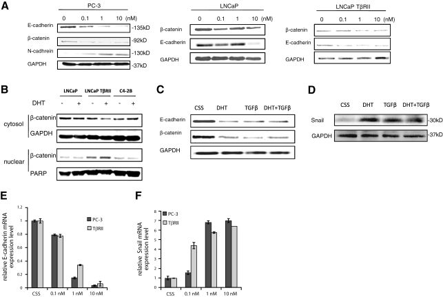
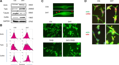
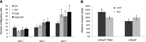
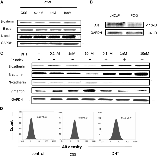
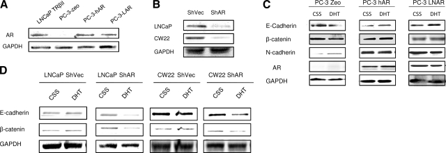
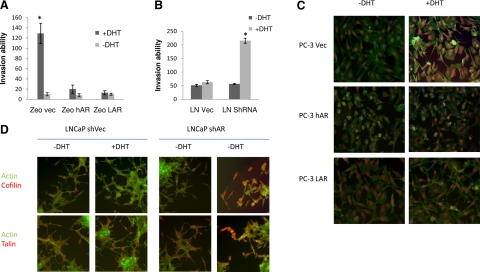
Androgens are functionally required for the normal growth of the prostate gland and in prostate tumor development and progression. Epithelial-mesenchymal-transition (EMT) is an important process during normal development and in cancer cell metastasis induced by factors within the microenvironment, such as transforming growth factor-beta (TGF-beta). This study examined the ability of androgens to influence EMT of prostate cancer epithelial cells. The EMT pattern was evaluated on the basis of expression of the epithelial markers E-cadherin/beta-catenin, and the mesenchymal markers N-cadherin, as well as cytoskeleton reorganization in response to 5alpha-dihydrotestosterone (DHT; 1 nM) and/or TGF-beta (5 ng/ml). Overexpressing and silencing approaches to regulate androgen receptor (AR) expression were conducted to determine the involvement of AR in EMT in the presence or absence of an AR antagonist. Our results demonstrate that androgens induce the EMT pattern in prostate tumor epithelial cell with Snail activation and lead to significant changes in prostate cancer cell migration and invasion potential. Expression levels of AR inversely correlated with androgen-mediated EMT in prostate tumor epithelial cells, pointing to a low AR content required for the EMT phenotype. These findings indicate the ability of androgens to induce EMT by potentially bypassing the functional involvement of TGF-beta, thus contributing to metastatic behavior of prostate cancer cells.-Zhum, M.-L., Kyprianou, N. Role of androgens and the androgen receptor in epithelial-mesenchymal transition and invasion of prostate cancer cells.
Figures
Similar articles
-
Profiling Prostate Cancer Therapeutic Resistance.Int J Mol Sci. 2018 Mar 19;19(3):904. doi: 10.3390/ijms19030904. Int J Mol Sci. 2018. PMID: 29562686 Free PMC article. Review.
-
TGF-beta signaling and androgen receptor status determine apoptotic cross-talk in human prostate cancer cells.Prostate. 2008 Feb 15;68(3):287-95. doi: 10.1002/pros.20698. Prostate. 2008. PMID: 18163430
-
Androgen receptor as a regulator of ZEB2 expression and its implications in epithelial-to-mesenchymal transition in prostate cancer.Endocr Relat Cancer. 2014 May 8;21(3):473-86. doi: 10.1530/ERC-13-0514. Print 2014 Jun. Endocr Relat Cancer. 2014. PMID: 24812058
-
Crosstalk between epithelial-mesenchymal transition and castration resistance mediated by Twist1/AR signaling in prostate cancer.Endocr Relat Cancer. 2015 Dec;22(6):889-900. doi: 10.1530/ERC-15-0225. Epub 2015 Aug 26. Endocr Relat Cancer. 2015. PMID: 26311513
-
Cell death under epithelial-mesenchymal transition control in prostate cancer therapeutic response.Int J Urol. 2018 Apr;25(4):318-326. doi: 10.1111/iju.13505. Epub 2018 Jan 17. Int J Urol. 2018. PMID: 29345000 Review.
Cited by
-
A novel HIF-1α-integrin-linked kinase regulatory loop that facilitates hypoxia-induced HIF-1α expression and epithelial-mesenchymal transition in cancer cells.Oncotarget. 2015 Apr 10;6(10):8271-85. doi: 10.18632/oncotarget.3186. Oncotarget. 2015. PMID: 25821081 Free PMC article.
-
The osteoblastic and osteoclastic interactions in spinal metastases secondary to prostate cancer.Cancer Growth Metastasis. 2013 Nov 27;6:61-80. doi: 10.4137/CGM.S12769. eCollection 2013. Cancer Growth Metastasis. 2013. PMID: 24665208 Free PMC article. Review.
-
Profiling Prostate Cancer Therapeutic Resistance.Int J Mol Sci. 2018 Mar 19;19(3):904. doi: 10.3390/ijms19030904. Int J Mol Sci. 2018. PMID: 29562686 Free PMC article. Review.
-
Novel pharmacologic targeting of tight junctions and focal adhesions in prostate cancer cells.PLoS One. 2014 Jan 31;9(1):e86238. doi: 10.1371/journal.pone.0086238. eCollection 2014. PLoS One. 2014. PMID: 24497940 Free PMC article.
-
Epithelial-mesenchymal transition in prostate cancer: an overview.Oncotarget. 2017 May 23;8(21):35376-35389. doi: 10.18632/oncotarget.15686. Oncotarget. 2017. PMID: 28430640 Free PMC article. Review.
References
-
- Jemal A, Siegel R, Ward E, Hao Y, Xu J, Murray T, Thun M J. Cancer statistics, 2008. CA Cancer J Clin. 2008;58:71–96. - PubMed
-
- Wang X, Yin L, Rao P, Stein R, Harsch K M, Lee Z, Heston W D. Targeted treatment of prostate cancer. J Cell Biochem. 2007;102:571–579. - PubMed
-
- Siiteri P K, Wilson J D. Testosterone formation and metabolism during male sexual differentiation in the human embryo. J Clin Endocrinol Metab. 1974;38:113–125. - PubMed
-
- Heinlein C A, Chang C. The roles of androgen receptors and androgen-binding proteins in nongenomic androgen actions. Mol Endocrinol. 2002;16:2181–2187. - PubMed
-
- Imperato-McGinley J, Binienda Z, Arthur A, Mininberg D T, Vaughan E D, Jr, Quimby F W. The development of a male pseudohermaphroditic rat using an inhibitor of the enzyme 5 alpha-reductase. Endocrinology. 1985;116:807–812. - PubMed
Publication types
MeSH terms
Substances
Grants and funding
LinkOut - more resources
Full Text Sources
Other Literature Sources
Medical
Research Materials
