. 2010 Jan 15;285(3):2064-76.
doi: 10.1074/jbc.M109.060988. Epub 2009 Nov 9.

Members of a novel protein family containing microneme adhesive repeat domains act as sialic acid-binding lectins during host cell invasion by apicomplexan parasites

Affiliations

Members of a novel protein family containing microneme adhesive repeat domains act as sialic acid-binding lectins during host cell invasion by apicomplexan parasites

Nikolas Friedrich et al. J Biol Chem. .

Abstract

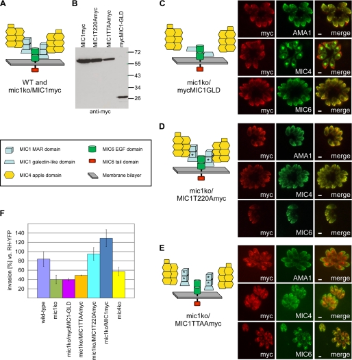
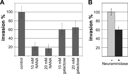
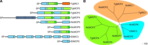
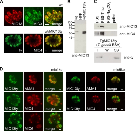
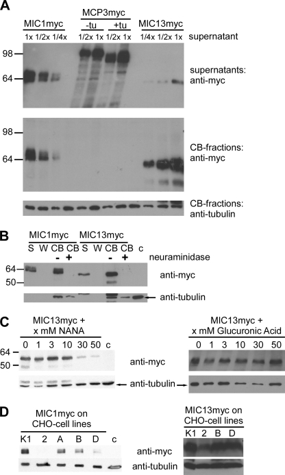
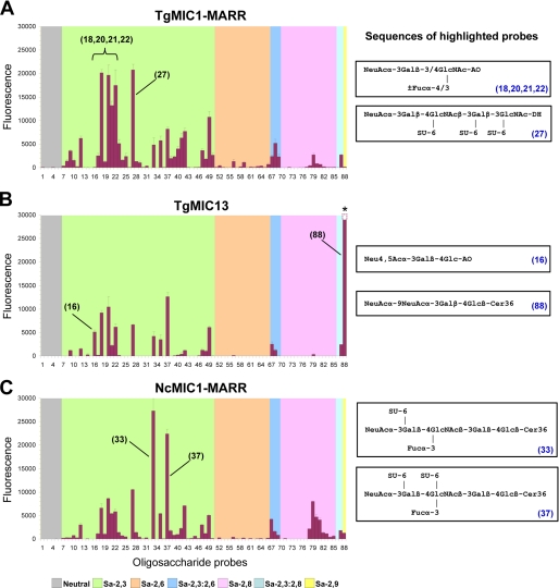
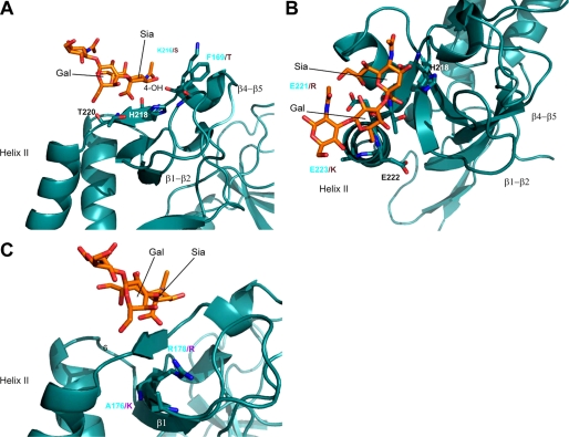
Numerous intracellular pathogens exploit cell surface glycoconjugates for host cell recognition and entry. Unlike bacteria and viruses, Toxoplasma gondii and other parasites of the phylum Apicomplexa actively invade host cells, and this process critically depends on adhesins (microneme proteins) released onto the parasite surface from intracellular organelles called micronemes (MIC). The microneme adhesive repeat (MAR) domain of T. gondii MIC1 (TgMIC1) recognizes sialic acid (Sia), a key determinant on the host cell surface for invasion by this pathogen. By complementation and invasion assays, we demonstrate that TgMIC1 is one important player in Sia-dependent invasion and that another novel Sia-binding lectin, designated TgMIC13, is also involved. Using BLAST searches, we identify a family of MAR-containing proteins in enteroparasitic coccidians, a subclass of apicomplexans, including T. gondii, suggesting that all these parasites exploit sialylated glycoconjugates on host cells as determinants for enteric invasion. Furthermore, this protein family might provide a basis for the broad host cell range observed for coccidians that form tissue cysts during chronic infection. Carbohydrate microarray analyses, corroborated by structural considerations, show that TgMIC13, TgMIC1, and its homologue Neospora caninum MIC1 (NcMIC1) share a preference for alpha2-3- over alpha2-6-linked sialyl-N-acetyllactosamine sequences. However, the three lectins also display differences in binding preferences. Intense binding of TgMIC13 to alpha2-9-linked disialyl sequence reported on embryonal cells and relatively strong binding to 4-O-acetylated-Sia found on gut epithelium and binding of NcMIC1 to 6'sulfo-sialyl Lewis(x) might have implications for tissue tropism.


Figures

FIGURE 1.
FIGURE 1.
Cooperative function for the Sia-binding TgMIC1-MAR domains and TgMIC4 in host cell invasion by T. gondii. A, schematic summarizing the domain organization of the components of the TgMIC1-4-6 complex in the WT and mic1ko/MIC1myc complemented strains. B, Western blot analysis of parasite lines expressing TgMIC1 mutant proteins on the mic1ko background. C–E, TgMIC1 mutant proteins expressed on the mic1ko background were assessed for their ability to substitute for TgMIC1 and target the components of the TgMIC1-4-6 complex to the micronemes. IFA was performed on intracellular parasites multiplying in their vacuole. TgAMA-1 is used as a micronemal marker independent of the TgMIC1-4-6 complex. Scale bars, 1 μm. A schematic summarizes the association/dissociation of the components of the TgMIC1-4-6 complex in the different strains. An asterisk indicates a Thr to Ala substitution in the Sia-binding site of the MAR domain. F, comparison of host cell invasion efficiency by the various T. gondii mutant strains using an RH-2YFP strain as internal standard for parasite fitness. Error bars, standard deviation.
FIGURE 2.
FIGURE 2.
Cell invasion assays using the T. gondii mic1ko strain demonstrate the existence of at least one more Sia-specific parasite lectin. A, invasion by mic1ko parasites in the absence and presence of 10 and 20 mm free NANA or galactose. B, assay comparing invasion of mic1ko parasites into host cells (HFFs) pretreated or not with neuraminidase. Error bars, standard deviation. Note that, compared with the parental strain, the mic1ko shows a 50% reduced invasion phenotype (Fig. 1F), but its invasion efficiency was set to 100% here.
FIGURE 3.
FIGURE 3.
Family of MAR domain-containing proteins in apicomplexans. A, schematic of the domain organization of various MCPs. MAR domain type I (light blue), MAR domain type II (light green), MAR domain type II extension the “β-finger” (green), galectin-like domain (orange), and region of short repeats (blue stripes) are shown. The presence of a Thr in a MAR domain in an equivalent position to those critical for Sia binding in TgMIC1-MARR is indicated by a black T. The fourth MAR domain in TgMCP4 contains this Thr, but the sequence context does not fit with it being indicative of a potential Sia-binding site; therefore, the T is in parentheses. B, phylogenetic relationship of MCPs from T. gondii, N. caninum, and E. tenella. Because the domain structure of the different proteins varies, only the sequence corresponding to the first two predicted MAR domains of each protein was used for the analysis. All bootstrap values are >80. Sequences were aligned in ClustalX, and alignment positions containing gaps in >50% of the sequences were excluded from phylogenetic analyses. Phylogenetic analyses were carried out using POWER (neighbor-joining distance method, bootstrapping with 1000 replicates). Phylogenetic trees were generated using TREEVIEW.
FIGURE 4.
FIGURE 4.
Characterization of TgMIC13 (TgMCP2) in T. gondii. A, IFA shows co-localization of TgMIC13 with microneme proteins in wild-type (wt) (top, confocal images) and in WT/TgMIC13ty parasites (bottom). B, Western blot analysis of endogenous and epitope-tagged TgMIC13 in wild-type and WT/TgMIC13ty parasites. An extract of HFF cells was also loaded as a control. C, assessment of TgMIC13 solubility by fractionation (top). Cell binding assays using ESA from WT/TgMIC13ty parasites (bottom). I, input; W, last of four washes; CB, cell-bound fraction. D, transient transfection of TgMIC13ty into the mic1ko and the mic6ko show correct localization of TgMIC13ty to the micronemes. Scale bars, 1 μm.
FIGURE 5.
FIGURE 5.
Binding characteristics of recombinant TgMIC13. A, cell binding assays comparing recombinant TgMIC1myc, TgMIC13myc, and α-factor-TgMCP3myc fusion protein produced in P. pastoris; +tu/−tu, protein expressed in the presence/absence of tunicamycin. Top panel, Western blot indicating the relative concentration of the proteins in supernatants used for the assay. Middle panel, cell-bound fractions (CB) probed for bound protein. Bottom panel, control for use of equivalent amounts of cell material in each experiment. B, pretreatment of cells with neuraminidase abolishes binding by TgMIC1 and TgMIC13. S, supernatant; W, last of four washes; CB, cell-bound fraction; c, background control. C, binding of TgMIC13 is strongly reduced in cell binding assays by competition with free NANA but not glucuronic acid. D, binding of TgMIC1 and TgMIC13 to various CHO cell lines (K1, WT strain; 2, lec2 mutant with strong reduction in Sia surface expression; A and B, pgsA745 and pgsB618 mutants deficient in glycosaminoglycan synthesis; D, pgsD677 mutant deficient in two glycosyltransferase activities). Only the cell-bound fractions of the assay are shown.
FIGURE 6.
FIGURE 6.
Carbohydrate microarray analyses of recombinant TgMIC1-MARR expressed in E. coli (A), TgMIC13 expressed in P. pastoris (B), and NcMIC1-MARR expressed in E. coli (C) using 88 lipid-linked oligosaccharide probes. Numerical scores of the binding signals are means of duplicate spots at 5 fmol/spot (with error bars). The complete list of probes and their sequences and binding scores are in supplemental Table 1. The binding signal for probe 88 was saturated and could not be accurately quantified (asterisk in B).
FIGURE 7.
FIGURE 7.
Three orientations of the crystal structure of TgMIC1-MARR in complex with 3′SiaLacNAc1–3 (Protein Data Bank code 3F5A) (29); 3′SiaLacNAc1–3 is shown in an orange stick representation. Key side chains are drawn as stick representations minus attached protons and labeled with sequence position. A and B, amino acid differences between TgMIC1 and TgMIC13 relevant to their binding specificities are labeled in cyan and violet, respectively. C, amino acid differences between TgMIC1 and NcMIC1 relevant to their binding specificities are labeled in cyan and violet, respectively.

Similar articles

Cited by

References

    1. Blumenschein T. M., Friedrich N., Childs R. A., Saouros S., Carpenter E. P., Campanero-Rhodes M. A., Simpson P., Chai W., Koutroukides T., Blackman M. J., Feizi T., Soldati-Favre D., Matthews S. (2007) EMBO J. 26, 2808–2820 - PMC - PubMed
    1. Persson K. E., McCallum F. J., Reiling L., Lister N. A., Stubbs J., Cowman A. F., Marsh K., Beeson J. G. (2008) J. Clin. Invest. 118, 342–351 - PMC - PubMed
    1. Sim B. K., Orlandi P. A., Haynes J. D., Klotz F. W., Carter J. M., Camus D., Zegans M. E., Chulay J. D. (1990) J. Cell Biol. 111, 1877–1884 - PMC - PubMed
    1. Takabatake N., Okamura M., Yokoyama N., Ikehara Y., Akimitsu N., Arimitsu N., Hamamoto H., Sekimizu K., Suzuki H., Igarashi I. (2007) Vet. Parasitol. 148, 93–101 - PubMed
    1. Martin M. J., Rayner J. C., Gagneux P., Barnwell J. W., Varki A. (2005) Proc. Natl. Acad. Sci. U.S.A. 102, 12819–12824 - PMC - PubMed

Publication types

MeSH terms