. 2010 Jan 1;285(1):793-804.
doi: 10.1074/jbc.M109.070037. Epub 2009 Nov 9.

Transcription factor GATA4 inhibits doxorubicin-induced autophagy and cardiomyocyte death

Affiliations

Transcription factor GATA4 inhibits doxorubicin-induced autophagy and cardiomyocyte death

Satoru Kobayashi et al. J Biol Chem. .

Abstract

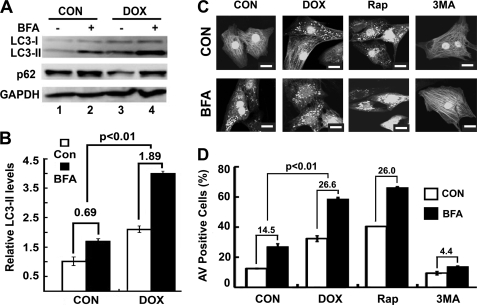
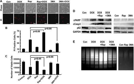
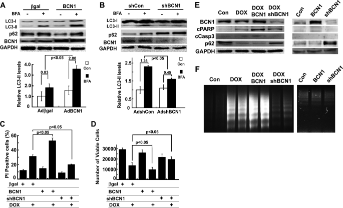
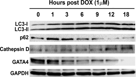
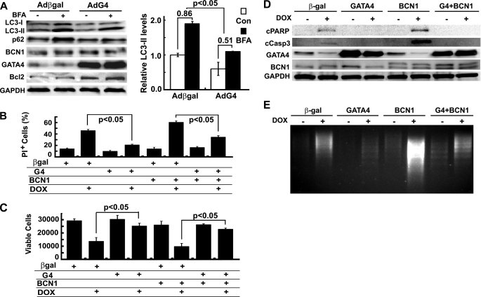
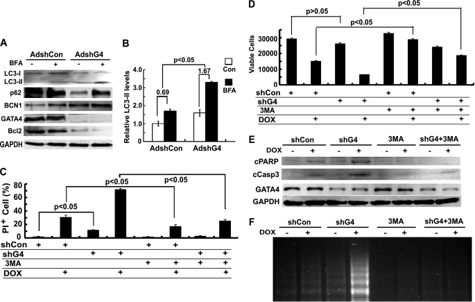
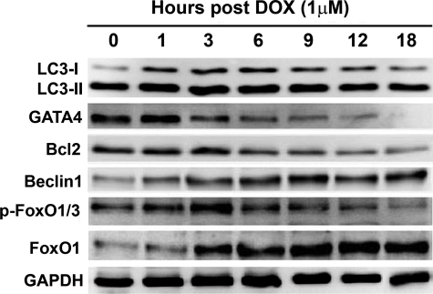
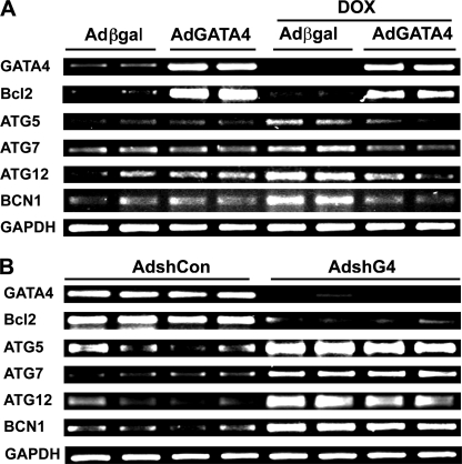
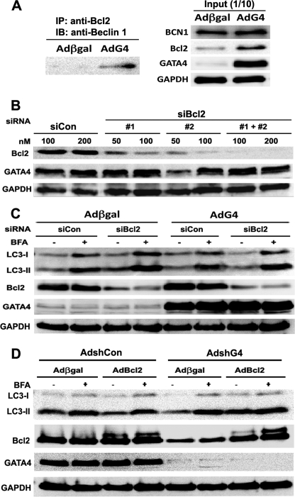
Doxorubicin (DOX) is a potent anti-tumor drug known to cause heart failure. The transcription factor GATA4 antagonizes DOX-induced cardiotoxicity. However, the protective mechanism remains obscure. Autophagy is the primary cellular pathway for lysosomal degradation of long-lived proteins and organelles, and its activation could be either protective or detrimental depending on specific pathophysiological conditions. Here we investigated the ability of GATA4 to inhibit autophagy as a potential mechanism underlying its protection against DOX toxicity in cultured neonatal rat cardiomyocytes. DOX markedly increased autophagic flux in cardiomyocytes as indicated by the difference in protein levels of LC3-II (microtubule-associated protein light chain 3 form 2) or numbers of autophagic vacuoles in the absence and presence of the lysosomal inhibitor bafilomycin A1. DOX-induced cardiomyocyte death determined by multiple assays was aggravated by a drug or genetic approach that activates autophagy, but it was attenuated by manipulations that inhibit autophagy, suggesting that autophagy contributes to DOX cardiotoxicity. DOX treatment depleted GATA4 protein levels, which predisposed cardiomyocytes to DOX toxicity. Indeed, GATA4 gene silencing triggered autophagy that rendered DOX more toxic, whereas GATA4 overexpression inhibited DOX-induced autophagy, reducing cardiomyocyte death. Mechanistically, GATA4 up-regulated gene expression of the survival factor Bcl2 and suppressed DOX-induced activation of autophagy-related genes, which may likely be responsible for the anti-apoptotic and anti-autophagic effects of GATA4. Together, these findings suggest that activation of autophagy mediates DOX cardiotoxicity, and preservation of GATA4 attenuates DOX cardiotoxicity by inhibiting autophagy through modulation of the expression of Bcl2 and autophagy-related genes.


Figures

FIGURE 1.
FIGURE 1.
DOX induced autophagy in cardiomyocytes. Neonatal rat cardiomyocytes were cultured in DMEM with 1% bovine serum and treated with DOX (1 μm) for 18 h. A, Western blots show protein levels of LC3-II and p62 in the absence and presence of the lysosome inhibitor BFA (50 nm). B, DOX increased autophagic flux (DOX 1.89 ± 0.02 versus control (CON) 0.69 ± 0.10), which is defined by the difference in LC3-II protein levels in the absence and presence of BFA. Data are expressed as the mean ± S.E. LC3-II levels were analyzed by 2-way ANOVA, and autophagic flux was analyzed by paired Student's t tests (n = 4). C, DOX induced accumulation of AVs. Cardiomyocytes were infected with AdGFP-LC3 and treated with DOX or saline in the absence or presence of BFA. Upon DOX treatment, GFP-LC3 formed punctate structures or dots, indicative of AVs. The green fluorescent images were converted to black/white for clarity. The scale bar is 20 μm. D, DOX increased autophagic flux as calculated by AV-positive cells with and without BFA (DOX 26.6 ± 1.9 versus CON 14.5 ± 0.4). The autophagic flux is the difference in the number of AV positive cells in the absence and presence of BFA. The AV positive cells were expressed as the percentage of cells that have >40 GFP-LC3 dots over the number of total GFP-expressing cells examined. Roughly 200 cells/6-cm plate were counted. Data were expressed as the mean ± S.E., and autophagic flux was analyzed by paired Student's t tests (n = 3). Rap and 3-MA were used as known autophagy inducer and inhibitor, respectively.
FIGURE 2.
FIGURE 2.
Rap enhanced DOX toxicity, whereas 3-MA attenuated it. Cardiomyocyte were treated with DOX and Rap (100 nm) or 3-MA (2.5 mm) for 18 h, and cardiomyocyte death was determined by PI staining (A and B), MTT assay (C), cleavage of PARP (cPARP) and caspase 3 (cCasp3, D), and DNA laddering (E). Data in B and C were expressed as mean ± S.E. and analyzed by one-way ANOVA (n = 3). The scale bars in A represent 200 μm.
FIGURE 3.
FIGURE 3.
Increased Beclin 1 (BCN1) aggravated DOX toxicity, but knockdown BCN1 reduced it. Cardiomyocytes were infected with AdBCN or AdshBCN for 24 or 48 h and then treated with DOX (1 μm) for another 18 h. Increased Beclin 1 expression accelerated autophagic flux (A, BCN1 2.0 ± 0.34 versus β-galactosidase 0.83 ± 0.30), whereas knocking down Beclin 1 reduced it (B, shBCN1 0.45 ± 0.33 versus shCon 1.34 ± 0.23), as indicated by the difference in protein levels of LC3-II in the absence and presence of BFA. Densitometry data were the mean ± S.E. (n = 6 for BCN1 and 3 for shBCN1) and were analyzed with two-way ANOVA followed by paired Student's t tests. Beclin 1 overexpression exacerbated DOX-induced cell death, and knocking down Beclin 1 reduced it, as determined by PI staining (C), MTT assay (D), cleavage of PARP (cPARP) and caspase 3 (cCasp3, E), and DNA laddering (F). Data in C and D were expressed as the mean ± S.E. and were analyzed by one-way ANOVA (n = 6 for BCN and 3 for shBCN1).
FIGURE 4.
FIGURE 4.
DOX-induced autophagy was associated with depleted protein levels of GATA4. Cardiomyocytes were treated with DOX for different time periods and then harvested for Western blot analysis. The increase in LC3-II levels was temporally linked with reduced p62 and diminished GATA4 protein levels.
FIGURE 5.
FIGURE 5.
Overexpression of GATA4 inhibited DOX-induced autophagy and cardiomyocyte death. Cardiomyocytes were infected with Adβgal or AdG4 and then treated with DOX for 18 h. A, Western blots show LC3-II and p62 levels (left), and a bar graph demonstrates autophagic flux index (β-galactosidase (β-gal) 0.86 ± 0.12 versus G4 0.51 ± 0.14) as indicated by the difference in protein levels of LC3-II in the absence and presence of BFA (right). Densitometry data were the mean ± S.E. (n = 3) and were analyzed with two-way ANOVA followed by paired Student's t tests. Cardiomyocyte death was determined by PI staining (B), MTT assay (C), cleavage of PARP (cPARP) and caspase 3 (cCasp3, D), and DNA laddering (E). Data in B and C were expressed as the mean ± S.E. and analyzed by one-way ANOVA (n = 3). Con, control.
FIGURE 6.
FIGURE 6.
Knocking down GATA4 triggered autophagy and exacerbated DOX-induced cardiomyocyte death. Cardiomyocytes were infected with AdshG4 or AdshCon for 48 h and then treated with DOX for 18 h. A, Western blots show LC3-II and p62 protein levels. B, shown is quantification of autophagic flux index (shCon 0.69 ± 0.26 versus shG4 1.67 ± 0.19) as determined by the difference in protein levels of LC3-II in the absence and presence of BFA. Densitometry data are the mean ± S.E. (n = 3) and were analyzed with two-way ANOVA followed by paired Student's t tests. Cardiomyocyte death was determined by PI staining (C), MTT assay (D), cleavage of PARP (cPARP) and caspase 3 (cCasp3, E), and DNA laddering (F). Data in C and D are expressed as the mean ± S.E. and were analyzed by one-way ANOVA (n = 3). Con, control.
FIGURE 7.
FIGURE 7.
DOX-induced elevation of LC3-II was temporally associated with depleted levels of GATA4 and Bcl2 but increased levels of Beclin 1 and FoxO1. Cardiomyocytes were treated with DOX for different time periods and then harvested for Western blot analysis.
FIGURE 8.
FIGURE 8.
GATA4 regulated the expression of Bcl2 and autophagy-related genes. A, cardiomyocytes were infected with Adβgal or AdG4 and then treated with DOX for 18 h. B, cardiomyocytes were infected with AdshG4 or AdshCon for 48 h. The mRNA expression of Bcl2 and several autophagy-related genes under both conditions were determined by semi-quantitative reverse transcriptase-PCR.
FIGURE 9.
FIGURE 9.
Bcl2 mediated the anti-autophagic effect of GATA4. A, increased GATA4 enhanced the interaction between Bcl2 and Beclin 1. Cardiomyocytes were infected with Adβgal or AdG4, and protein samples were prepared 24 h later. 100 μg of protein was used for co-IP with Bcl2 antibodies (left panel), and 10 μg of protein was used as the input control (right panel). IB, immunoblotting. B, two different siRNAs against rat Bcl2 mRNA sequence (siBcl2) were transfected into cardiomyocytes. Both siRNAs were able to knock down Bcl2 protein levels in a dose-dependent manner, with siBcl2 #2 having a higher efficiency. C, cardiomyocytes were infected with Adβgal or AdG4 and then followed by siRNA transfection with a Silencer Negative Control siRNA (siCon) or a mixture of the two siBcl2 (#1 and #2 each at 50 nm). Knocking down Bcl2 markedly attenuated the ability of GATA4 to inhibit autophagy as shown by the difference in protein levels of LC3-II in the absence and presence of BFA. D, cardiomyocytes were infected with AdshG4 or AdshCon for 48 h and then infected with AdBcl2 for another 24 h. Western blot analysis was performed showing that GATA4 depletion-induced autophagy was reversed by Bcl2 overexpression.

References

    1. Swain S. M., Whaley F. S., Ewer M. S. (2003) Cancer 97, 2869–2879 - PubMed
    1. Singal P. K., Iliskovic N. (1998) N. Engl. J. Med. 339, 900–905 - PubMed
    1. Minotti G., Menna P., Salvatorelli E., Cairo G., Gianni L. (2004) Pharmacol. Rev. 56, 185–229 - PubMed
    1. Kumar D., Kirshenbaum L. A., Li T., Danelisen I., Singal P. K. (2001) Antioxid. Redox Signal. 3, 135–145 - PubMed
    1. Yen H. C., Oberley T. D., Vichitbandha S., Ho Y. S., St Clair D. K. (1996) J. Clin. Invest. 98, 1253–1260 - PMC - PubMed

Publication types

MeSH terms

LinkOut - more resources