. 2010 Feb 5;285(6):3608-3616.
doi: 10.1074/jbc.M109.066431. Epub 2009 Nov 9.

SAFB1 mediates repression of immune regulators and apoptotic genes in breast cancer cells

Affiliations

SAFB1 mediates repression of immune regulators and apoptotic genes in breast cancer cells

Stephanie Hammerich-Hille et al. J Biol Chem. .

Abstract

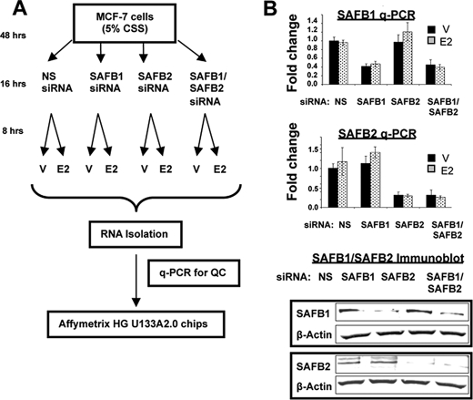
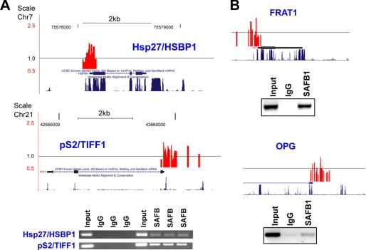
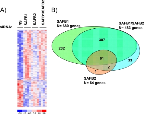
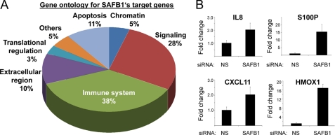
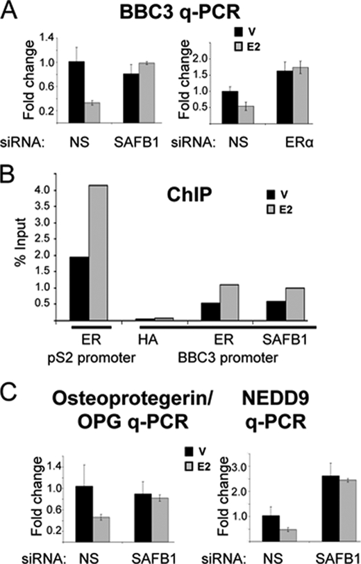
The scaffold attachment factors SAFB1 and SAFB2 are paralogs, which are involved in cell cycle regulation, apoptosis, differentiation, and stress response. They have been shown to function as estrogen receptor corepressors, and there is evidence for a role in breast tumorigenesis. To identify their endogenous target genes in MCF-7 breast cancer cells, we utilized a combined approach of chromatin immunoprecipitation (ChIP)-on-chip and gene expression array studies. By performing ChIP-on-chip on microarrays containing 24,000 promoters, we identified 541 SAFB1/SAFB2-binding sites in promoters of known genes, with significant enrichment on chromosomes 1 and 6. Gene expression analysis revealed that the majority of target genes were induced in the absence of SAFB1 or SAFB2 and less were repressed. Interestingly, there was no significant overlap between the genes identified by ChIP-on-chip and gene expression array analysis, suggesting regulation through regions outside the proximal promoters. In contrast to SAFB2, which shared most of its target genes with SAFB1, SAFB1 had many unique target genes, most of them involved in the regulation of the immune system. A subsequent analysis of the estrogen treatment group revealed that 12% of estrogen-regulated genes were dependent on SAFB1, with the majority being estrogen-repressed genes. These were primarily genes involved in apoptosis, such as BBC3, NEDD9, and OPG. Thus, this study confirms the primary role of SAFB1/SAFB2 as corepressors and also uncovers a previously unknown role for SAFB1 in the regulation of immune genes and in estrogen-mediated repression of genes.


Figures

FIGURE 1.
FIGURE 1.
Design of gene expression array study, and quality control of SAFB1/SAFB2 siRNA in MCF-7 cells. A, model of experimental setup of gene expression array study. V, vehicle. B, specific and efficient down-regulation of SAFB1 and SAFB2 by siRNA. MCF-7 cells were transiently transfected with NS siRNA, SAFB1 siRNA, SAFB2 siRNA, or SAFB1/SAFB2 siRNA. mRNA levels of SAFB1 and SAFB2 were measured by q-PCR (top panel), and protein expression by immunoblotting (bottom panel) using antibodies as indicated.
FIGURE 2.
FIGURE 2.
Confirmation of SAFB binding to candidate genes identified in ChIP-chip study. A, confirmation of SAFB binding to control genes hsp27/HSPB1 and pS2/TIFF1. Top panel, graphical presentation of SAFB1-binding peaks in hsp27/HSPB1 and pS2/TIFF1 genes, as identified in the ChIP-chip experiment. The black line around 1 represent the average binding (signal) across the entire array. The gene structure and the mammalian conservation of sequence are shown. Bottom panel, ChIP was performed using pan-SAFB antibodies and triplicate biological samples (primer sequences are provided in supplemental Table S1). B, same as in A, but with FRAT1 and OPG genes. ChIP assay, using SAFB1-specific antibodies, is representative of three independent experiments.
FIGURE 3.
FIGURE 3.
Identification of SAFB1, SAFB2, and SAFB1/SAFB2 target genes in MCF-7 cells. A, heat map representing 716 genes that were significantly regulated by SAFB1, SAFB2, or the combination of SAFB1/SAFB2 siRNA. The cutoff for this analysis was based on a Benjamin-Hochberg adjusted p = 0.1. Expression levels are shown in red and blue (levels are above and below the median, respectively). B, SAFB1 regulates more target genes than SAFB2. The Venn diagram represents genes that are significantly regulated by SAFB1, SAFB2, and SAFB1/SAFB2.
FIGURE 4.
FIGURE 4.
SAFB1 regulates immune genes. A, gene ontology analysis for SAFB1-regulated genes (n = 680) revealed that the majority represent immune regulatory, signaling, and apoptotic genes. B, SAFB1 is necessary for the repression of immune regulatory genes. The SAFB1-mediated repression of four immune regulatory SAFB1 candidate genes was tested (IL8, S100P, CXCL11, and HMOX1). q-PCR was performed using RNA from MCF-7 cells transfected with NS siRNA or SAFB1 siRNA. The data are an average of three replicates ± S.D.
FIGURE 5.
FIGURE 5.
SAFB1 is involved in estrogen-mediated repression of apoptotic genes. A, E2 repression of BBC3 is SAFB1 and ERα-dependent. BBC3 q-PCR was performed using RNA from MCF-7 cells transfected with NS siRNA, SAFB1 siRNA (left panel), or ERα siRNA (right panel) and treated with vehicle (V) (black) or estrogen (gray) (8 h). The data are an average of three replicates ± S.D. B, ERα and SAFB1 are recruited to the BBC3 proximal promoter. Results of a ChIP analysis on the BBC3 promoter using antibodies against hemagglutinin (negative control), ERα, and SAFB1 are shown. For the ChIP, MCF-7 cells were treated with vehicle (black) or estrogen (gray) for 45 min. Recruitment of ERα to the pS2 promoter was used as positive control. C, loss of estrogen repression of osteoprotegerin and NEDD9 in the absence of SAFB1. The E2-mediated repression of osteoprotegerin and NEDD9 was tested using RNA from MCF-7 cells transfected with NS siRNA and SAFB1 siRNA and treated with vehicle (black) or estrogen (gray) (8 h). q-PCR analysis of mRNA was performed, and the data shown are an average of three replicates ± S.D.

Similar articles

Cited by

References

    1. Deroo B. J., Korach K. S. (2006) J. Clin. Invest. 116, 561–570 - PMC - PubMed
    1. O'Malley B. W. (2007) Mol. Endocrinol. 21, 1009–1013 - PubMed
    1. Acevedo M. L., Lee K. C., Stender J. D., Katzenellenbogen B. S., Kraus W. L. (2004) Mol. Cell 13, 725–738 - PubMed
    1. Dobrzycka K. M., Townson S. M., Jiang S., Oesterreich S. (2003) Endocr. Relat. Cancer 10, 517–536 - PubMed
    1. Oesterreich S., Zhang Q., Hopp T., Fuqua S. A., Michaelis M., Zhao H. H., Davie J. R., Osborne C. K., Lee A. V. (2000) Mol. Endocrinol. 14, 369–381 - PubMed

Publication types

MeSH terms

Associated data