. 2010 Jan;78(1):301-8.
doi: 10.1128/IAI.00959-09. Epub 2009 Nov 9.

CXCL10 production by human monocytes in response to Leishmania braziliensis infection

Affiliations

CXCL10 production by human monocytes in response to Leishmania braziliensis infection

Diego A Vargas-Inchaustegui et al. Infect Immun. 2010 Jan.

Abstract

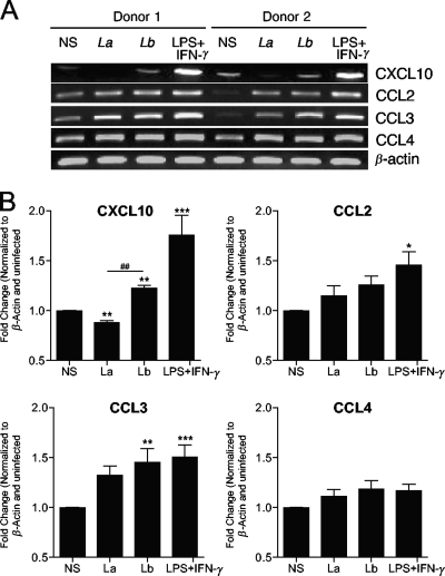
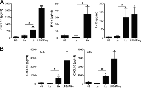
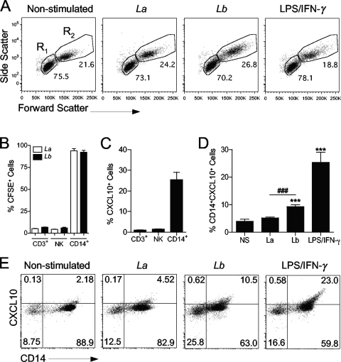
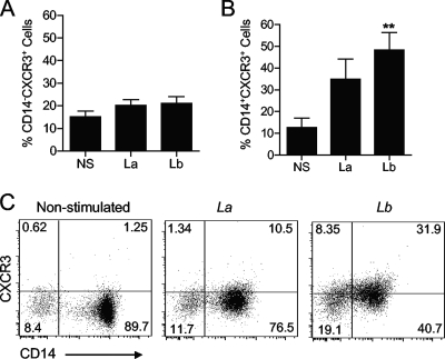
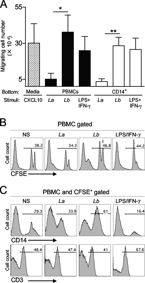
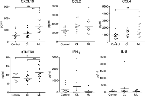
Leishmania (subgenus Viannia) braziliensis is the causative agent of mucocutaneous leishmaniasis (ML) in South America, and ML is characterized by excessive T- and B-cell responses to the parasite. We speculate that the unbalanced production of inflammatory mediators in response to L. braziliensis infection contributes to cell recruitment and disease severity. To test this hypothesis, we first examined the response of peripheral blood mononuclear cells (PBMCs) from healthy volunteers to L. braziliensis infection. We observed that while L. braziliensis infection induced the production of chemokine (C-X-C motif) ligand 10 (CXCL10) and interleukin-10 (IL-10) in human PBMCs and macrophages (MPhis), enhanced expression of CXCL10 and its receptor, chemokine CXC receptor (CXCR3), was predominantly detected in CD14(+) monocytes. The chemoattractant factors secreted by L. braziliensis-infected cells were highly efficient in recruiting uninfected PBMCs (predominantly CD14(+) cells) through Transwell membranes. Serum samples from American tegumentary leishmaniasis (ATL) patients (especially the ML cases) had significantly higher levels of CXCL10, CCL4, and soluble tumor necrosis factor (TNF) receptor II (sTNFRII) than did those of control subjects. Our results suggest that, following L. braziliensis infection, the production of multiple inflammatory mediators by the host may contribute to disease severity by increasing cellular recruitment.


Figures

FIG. 1.
FIG. 1.
Activation of chemokine gene transcription by human PBMCs in response to L. braziliensis infection. Human PBMCs were isolated from healthy donors (n = 8) and infected with L. braziliensis (Lb and Lb) or L. amazonensis (La and La) promastigotes at a 5:1 parasite-to-cell ratio. At 4 h p.i, total RNA was extracted for RT-PCR analysis. Human PBMCs were isolated from nonstimulated controls (NS). LPS plus IFN-γ were used as a positive control. (A) Gel images of CXCL10, CCL2, CCL3, and CCL4 transcripts from two donors. (B) Densitometry data obtained from all donors were pooled and shown in the plots. represents Values that are statistically significantly different (P < 0.001) between the compared groups are indicated (##). Values that are statistically significantly different between the nonstimulated control and infected groups are indicated as follows: *, P < 0.05; **, P < 0.01; ***, P < 0.001.
FIG. 2.
FIG. 2.
L. braziliensis infection induces CXCL10 production in human PBMCs and macrophages. (A) PBMCs were isolated from healthy donors (n = 10) and infected with L. braziliensis (Lb) or L. amazonensis (La) promastigotes at a 5:1 parasite-to-cell ratio for 24 h. (B) CD14+ monocytes were isolated from healthy donor PBMCs (n = 3) and differentiated into MΦs by cultivation in conditioned medium for 5 days. MΦs were infected with promastigotes at a 10:1 parasite-to-cell ratio for 24 and 48 h. Cells stimulated with LPS/IFN-γ (100 ng/ml each) were used as positive controls. The level of the indicated molecule in culture supernatants was assayed by ELISA. Values that are statistically significantly different between the compared groups are indicated as follows: #, P < 0.05; ##, P < 0.01. Values that are statistically significantly different between the nonstimulated control (NS) and infected groups are indicated as follows: *, P < 0.05; **, P < 0.01; ***, P < 0.001.
FIG. 3.
FIG. 3.
Monocytes are the major producers of CXCL10 following L. braziliensis infection. Human PBMCs were isolated from healthy donors (n = 6) and infected with CFSE-labeled L. braziliensis (Lb) or L. amazonensis (La) promastigotes at a 5:1 parasite-to-cell ratio for 24 h. (A) Representative forward scatter versus side scatter plots of PBMCs cultured under different conditions are shown. Gates used to analyze lymphocytes (R1) and monocytes (R2) are indicated. 50K, 50,000. (B) CFSE intensity was used to determine the infection rates among T lymphocytes (CD3+), NK cells (CD3 CD56+), and monocytes (CD14+ cells within the monocyte gate). (C) CXCL10 production in different cell types in response to LPS/IFN-γ (100 ng/ml each) stimulation was assayed by FACS. (D and E) Intracellular CXCL10 in infected monocytes (CD14+ CFSE+) was measured by FACS. Values that are statistically significantly different (P < 0.001) between the compared groups are indicated (###). Values that are statistically significantly different (P < 0.001) between the nonstimulated control and infected groups are indicated (***). Numbers shown in panel E represent the percentage of cells in each quadrant.
FIG. 4.
FIG. 4.
Monocyte-specific upregulation of CXCR3 after Leishmania infection. PBMCs were isolated from healthy donors (n = 6) and infected with L. braziliensis (Lb) or L. amazonensis (La) promastigotes at a 5:1 parasite-to-cell ratio for 24 h. The levels of CXCR3 expression on CD14 (A) and CD14+ cells (B and C) were measured by FACS. Values that are statistically significantly different (P < 0.01) between the nonstimulated control (NS) and infected groups are indicated (**). Numbers shown in panel C represent the percentage of cells in each quadrant.
FIG. 5.
FIG. 5.
L. braziliensis-infected PBMCs and monocytes efficiently induce migration of uninfected PBMCs. PBMCs and CD14+ cells were isolated from healthy donors (n = 4) and infected with L. braziliensis (Lb) or L. amazonensis (La) promastigotes at a 5:1 parasite-to-cell ratio and placed in the basal chambers of Transwell plates. Noninfected, donor-matched PBMCs were labeled with CFSE, placed in the apical chamber, and allowed to migrate in response to chemotactic factors released from the basal chamber for 14 h. Cells stimulated with LPS/IFN-γ (100 ng/ml each) were used as positive controls. (A) The total number of migrating cells present in the basal chamber was counted after the incubation period. Data were normalized by subtracting the migration observed in nonstimulated controls. Values that are statistically significantly different by one-way ANOVA are indicated as follows: *, P < 0.05; **, P < 0.01. (B) The percentage of migrating CFSE+ PBMCs in the basal chamber was measured by FACS. (C) Percentages of monocytes (CD14+) and T lymphocytes (CD3+) within CFSE+ migrating cells. (B and C) Histograms from one of four representative experiments are shown. NS, nonstimulated controls.
FIG. 6.
FIG. 6.
Profiles of inflammatory chemokines and cytokines in serum samples from American tegumentary leishmaniasis patients. Serum samples were collected from a total of 13 CL patients and 14 patients with active ML. Samples from 13 healthy volunteers were used as controls. The levels of the indicated molecules were measured with a RayBiotech custom Quantibody array. Each circle shows the value for an individual patient or healthy volunteer, and the short horizontal bar shows the mean value for that group. Values that are statistically significantly different between the compared groups by one-way ANOVA are indicated by bars and asterisks as follows: *, P < 0.05; **, P < 0.01; ***, P < 0.001.

Similar articles

Cited by

References

    1. Alexander, J., A. R. Satoskar, and D. G. Russell. 1999. Leishmania species: models of intracellular parasitism. J. Cell Sci. 112:2993-3002. - PubMed
    1. Amato, V. S., H. F. de Andrade, and M. I. Duarte. 2003. Mucosal leishmaniasis: in situ characterization of the host inflammatory response, before and after treatment. Acta Trop. 85:39-49. - PubMed
    1. Arevalo, J., L. Ramirez, V. Adaui, M. Zimic, G. Tulliano, C. Miranda-Verastegui, M. Lazo, R. Loayza-Muro, S. De Doncker, A. Maurer, F. Chappuis, J. C. Dujardin, and A. Llanos-Cuentas. 2007. Influence of Leishmania (Viannia) species on the response to antimonial treatment in patients with American tegumentary leishmaniasis. J. Infect. Dis. 195:1846-1851. - PubMed
    1. Asensio, V. C., J. Maier, R. Milner, K. Boztug, C. Kincaid, M. Moulard, C. Phillipson, K. Lindsley, T. Krucker, H. S. Fox, and I. L. Campbell. 2001. Interferon-independent, human immunodeficiency virus type 1 gp120-mediated induction of CXCL10/IP-10 gene expression by astrocytes in vivo and in vitro. J. Virol. 75:7067-7077. - PMC - PubMed
    1. Auffray, C., M. H. Sieweke, and F. Geissmann. 2009. Blood monocytes: development, heterogeneity, and relationship with dendritic cells. Annu. Rev. Immunol. 27:669-692. - PubMed

Publication types

MeSH terms