. 2010 Jan;30(2):372-81.
doi: 10.1128/MCB.00063-09. Epub 2009 Nov 9.

KLF4 interacts with beta-catenin/TCF4 and blocks p300/CBP recruitment by beta-catenin

Affiliations

KLF4 interacts with beta-catenin/TCF4 and blocks p300/CBP recruitment by beta-catenin

Paul M Evans et al. Mol Cell Biol. 2010 Jan.

Abstract

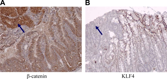
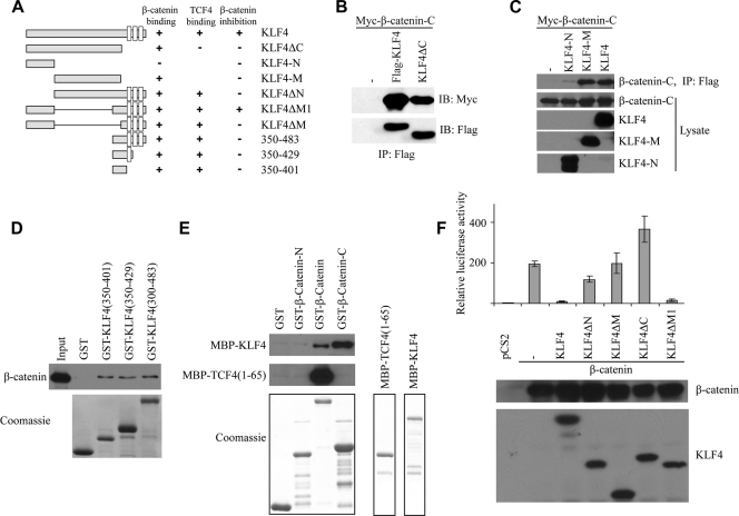
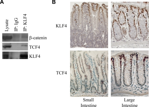
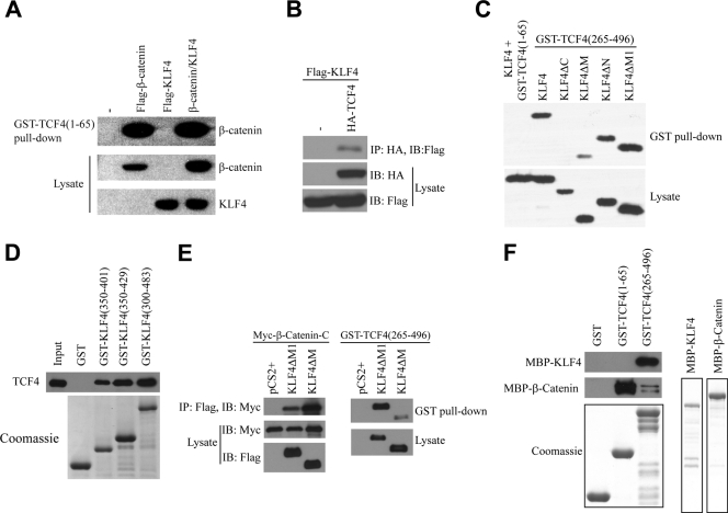
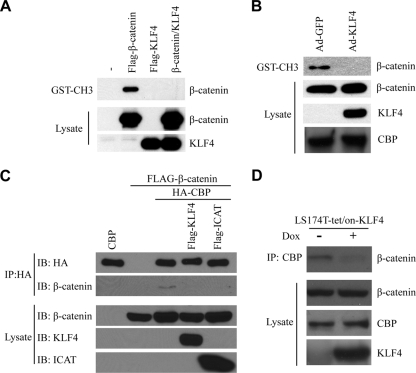
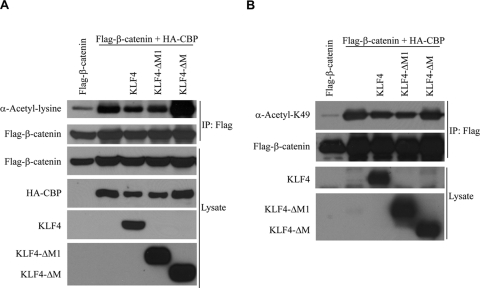
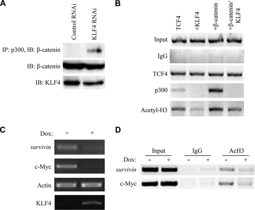
Wnt signaling is crucial in the organization and maintenance of the human intestinal epithelium, and somatic mutations that result in deregulated Wnt signaling are an early event in the development of colorectal cancer. The Wnt ligand ultimately results in the stabilization of cytoplasmic beta-catenin, which is then free to enter the nucleus and activate transcription through its interaction with the transcription factor TCF4. Our laboratory recently found that KLF4, a transcription factor highly expressed in the adult intestine and critical for intestinal differentiation, interacts with beta-catenin and inhibits Wnt signaling. In this study, we characterize the molecular mechanisms of KLF4-mediated inhibition of Wnt/beta-catenin signaling. We find that the KLF4 directly interacts with the C-terminal transactivation domain of beta-catenin and inhibits p300/CBP recruitment by beta-catenin. KLF4 inhibits p300/CBP-mediated beta-catenin acetylation as well as histone acetylation on Wnt target genes. In addition, we observe that KLF4 directly interacts with TCF4 independently of beta-catenin and that KLF4 and TCF4 are expressed in similar patterns within the large intestine, with greatest staining near the epithelial surface. These results provide a deeper understanding of the regulation of beta-catenin in the intestine and will have important implications in cancer and stem cell research.


Figures

FIG. 1.
FIG. 1.
KLF4 expression in the intestine of the ApcMin/+ mouse. (A) β-Catenin levels are increased in the nuclei of adenomas (arrow) compared with their levels in adjacent normal intestine. (B) KLF4 levels are decreased in adenomas (arrow) compared with their levels in adjacent normal intestine.
FIG. 2.
FIG. 2.
The N-terminal and C-terminal domains of KLF4 are required for inhibition of the TOPFlash reporter. (A) Diagram of all KLF4 constructs used in this paper. All constructs contain an N-terminal Flag tag. Full-length KLF4 contains 483 amino acids. The open boxes shown in the C termini represent zinc finger motifs. The binding affinity and activity of each construct are summarized in adjacent columns. (B) HEK293T cells were transfected with Myc-tagged β-catenin C-terminal transactivation domain and full-length KLF4 or KLF4ΔC (deletion of C-terminal zinc fingers). Anti-Flag immunoprecipitation was performed, followed by anti-Myc Western blotting. (C) HEK293T cells were transfected with a Myc-tagged β-catenin C terminus and either the N-terminal (amino acids 1 to 157) or middle (amino acids 157 to 401) domain of the full-length KLF4 protein. After anti-Flag immunoprecipitation, a Myc Western blot assay was performed (top row). Western blotting was performed on cell lysate as a control. (D) SW480 cell lysate was incubated with various GST-tagged KLF4 constructs as shown. After several washings, bound protein was eluted and an anti-β-catenin Western blot assay was performed. Coomassie staining demonstrates the expression level of each GST-tagged protein (lower panel). (E) KLF4 directly interacts with β-catenin. The GST-tagged β-catenin N terminus, β-catenin, and β-catenin C terminus were incubated with MBP-tagged KLF4 or TCF4(1-65). β-Catenin proteins were pulled down with GST beads, and KLF4 and TCF4 were analyzed by Western blotting with an anti-MBP antibody. Coomassie staining demonstrates expression level of each purified protein (lower panel). (F) HEK293T cells were plated in 12-well plates, and each well was transfected with 0.2 μg Super8xTOPFlash, 0.05 μg Renilla luciferase, 0.5 μg of β-catenin DNA, and 0.5 μg of one of the various KLF4 constructs shown. Cells were harvested 48 h later and analyzed for luciferase activity. Renilla was used as a control for transfection efficiency and used to normalize raw luciferase data. Data points were performed in triplicate, and values are expressed as fold increases relative to controls (empty vector only). Western blotting demonstrates similar levels of expression of β-catenin between conditions and of expression levels of various KLF4 constructs (lower panel).
FIG. 3.
FIG. 3.
KLF4 and TCF4 interact. (A) Endogenous KLF4 was immunoprecipitated from HCT116 cells, and Western blotting was performed on the eluate. Both β-catenin and TCF4 were detectable in the eluate. IgG was used as a negative control. (B) Immunohistochemistry of the mouse small and large intestine. Expression patterns of KLF4 and TCF4 in the mouse intestine overlap, with the strongest expression near the epithelial surface. Tissue was harvested from the small and large intestines of an adult mouse, embedded in paraffin, and stained using either a KLF4 or TCF4 antibody. The base of the crypts is at the bottom of each photo, whereas the tip of the villus (small intestine) or luminal surface (large intestine) is at the top of the photo.
FIG. 4.
FIG. 4.
The C-terminal domain of KLF4 and the DNA-binding domain of TCF4 interact. (A) HEK293T cells were transfected with Flag-β-catenin, Flag-KLF4, or both, and after 48 h, lysate was collected and incubated with GST-tagged TCF4(1-65). After several washings, bound proteins were eluted and an anti-Flag Western blot assay was performed. (B) HEK293T cells were transfected with Flag-tagged KLF4, with or without HA-tagged TCF4, lysate was harvested, and anti-HA immunoprecipitation was performed. An anti-Flag Western blot assay was then performed on the bound proteins. (C) GST-TCF4(1-65) or GST-TCF4(265-496) was incubated with lysate from HEK293T cells overexpressing various Flag-tagged KLF4 constructs as shown. After several washings, bound proteins were eluted and an anti-Flag Western blot assay was performed. (D) HEK293T cells were transfected with a Myc-tagged TCF4 construct. Forty-eight hours later, cell lysate was collected and incubated with various GST-tagged KLF4 constructs. After several washings, bound proteins were eluted and an anti-Myc Western blot assay was performed. Coomassie staining was performed to demonstrate expression levels of the various GST-tagged proteins used. (E) KLF4ΔM1 binds both β-catenin and TCF4. Left: the Myc-tagged β-catenin C terminus was cotransfected with Flag-tagged KLF4ΔM and KLF4ΔM1 into HEK293T cells. β-Catenin was immunoprecipitated with an anti-Myc antibody, and KLF4ΔM and KLF4ΔM1 were analyzed by Western blotting with an anti-Flag antibody. Right: Flag-tagged KLF4ΔM and KLF4ΔM1 were transfected into HEK293T cells and then incubated with GST-TCF4(265-496). TCF4 was pulled down with GST beads, and KLF4ΔM and KLF4ΔM1 were analyzed by Western blotting. (F) KLF4 directly interacts with TCF4. GST-tagged TCF4(1-65) and TCF4(265-496) were incubated with MBP-tagged KLF4 or β-catenin. TCF4 proteins were pulled down with GST beads, and KLF4 and β-catenin were analyzed by Western blotting with an anti-MBP antibody. Coomassie staining demonstrates the expression level of each purified protein (lower panel).
FIG. 5.
FIG. 5.
KLF4 inhibits binding between β-catenin and p300/CBP. (A) HEK293T cells were transfected with Flag-β-catenin, Flag-KLF4, or both, and after 48 h, lysate was collected and incubated with the GST-tagged CH3 domain of p300. After several washings, bound proteins were eluted and an anti-Flag Western blot assay was performed. (B) HCT116 cells were treated with either control adenovirus (Ad-GFP) or a KLF4-expressing adenovirus (Ad-KLF4). After 48 h, cell lysate was collected and incubated with the GST-tagged CH3 domain of p300. After several washings, bound proteins were eluted and an anti-β-catenin Western blot assay was performed. Western blotting against CBP demonstrates that overexpression of KLF4 did not affect the expression of CBP. (C) HEK293T cells were transfected with HA-tagged CBP and Flag-tagged β-catenin, with or without Flag-KLF4 or ICAT. Anti-HA immunoprecipitation was performed on the cell lysate, and bound proteins were analyzed via anti-Flag Western blotting. (D) Expression of KLF4 was induced in LS174T-tet/on-KLF4 cells via treatment with doxycycline for 48 h. Anti-CBP immunoprecipitation was performed on cell lysate, and bound proteins were analyzed via an anti-β-catenin Western blot assay.
FIG. 6.
FIG. 6.
KLF4 inhibits acetylation of β-catenin. (A) HEK293T cells were transfected with Flag-β-catenin, HA-CBP, and various Flag-tagged KLF4 constructs. After 48 h, anti-Flag immunoprecipitation was performed on cell lysate. After several washings, bound proteins were analyzed via an anti-acetylated lysine Western blot assay. (B) HEK293T cells were transfected with Flag-β-catenin, HA-CBP, and various Flag-tagged KLF4 constructs. After 48 h, cell lysate was collected and directly analyzed via an anti-acetylated lysine 49-β-catenin Western blot assay.
FIG. 7.
FIG. 7.
KLF4 inhibits β-catenin-mediated recruitment of p300/CBP. (A) HCT116 cells were transfected with either control RNAi or KLF4 RNAi, and after 48 h, anti-p300 immunoprecipitation was performed on the cell lysate. Bound proteins were washed and analyzed via anti-β-catenin Western blotting. (B) HEK293T cells were transfected with the Super8xTOPFlash plasmid as well as the expression plasmids shown. After 48 h, cells were cross-linked and sonicated, and immunoprecipitation was performed using the antibodies shown at left as row labels. Bound DNA-protein complexes were eluted and de-cross-linked, and free DNA was amplified using primers specific to the promoter region of the Super8xTOPFlash reporter. (C) Expression of KLF4 was induced in LS174T-tet/on-KLF4 cells via treatment with doxycycline for 48 h. Total RNA was then harvested, and cDNA was produced using reverse transcriptase and amplified via PCR using primers specific to the genes shown. Actin was included as a negative control. (D) Expression of KLF4 was induced in LS174T-tet/on-KLF4 cells via treatment with doxycycline for 48 h. After 48 h, cells were cross-linked and sonicated, and immunoprecipitation was performed using antibodies shown at left as row labels. Bound DNA-protein complexes were eluted and de-cross-linked, and free DNA was amplified using primers specific to the promoter region of the genes shown.

References

    1. Behrens, J., J. P. von Kries, M. Kuhl, L. Bruhn, D. Wedlich, R. Grosschedl, and W. Birchmeier. 1996. Functional interaction of beta-catenin with the transcription factor LEF-1. Nature 382:638-642. - PubMed
    1. Belenkaya, T. Y., C. Han, H. J. Standley, X. Lin, D. W. Houston, J. Heasman, and X. Lin. 2002. Pygopus encodes a nuclear protein essential for Wingless/Wnt signaling. Development 129:4089-4101. - PubMed
    1. Billin, A. N., H. Thirlwell, and D. E. Ayer. 2000. Beta-catenin-histone deacetylase interactions regulate the transition of LEF1 from a transcriptional repressor to an activator. Mol. Cell. Biol. 20:6882-6890. - PMC - PubMed
    1. Brannon, M., J. D. Brown, R. Bates, D. Kimelman, and R. T. Moon. 1999. XCtBP is a XTcf-3 co-repressor with roles throughout Xenopus development. Development 126:3159-3170. - PubMed
    1. Cavallo, R. A., R. T. Cox, M. M. Moline, J. Roose, G. A. Polevoy, H. Clevers, M. Peifer, and A. Bejsovec. 1998. Drosophila Tcf and Groucho interact to repress Wingless signalling activity. Nature 395:604-608. - PubMed

Publication types

LinkOut - more resources