

doi: 10.1128/MCB.00916-08.
Epub 2009 Nov 9.
Aurora-A phosphorylates, activates, and relocalizes the small GTPase RalA
Affiliations
- PMID: 19901077
- PMCID: PMC2798468
- DOI: 10.1128/MCB.00916-08
Item in Clipboard
Aurora-A phosphorylates, activates, and relocalizes the small GTPase RalA
Mol Cell Biol.
2010 Jan.
Abstract
The small GTPase Ras, which transmits extracellular signals to the cell, and the kinase Aurora-A, which promotes proper mitosis, can both be inappropriately activated in human tumors. Here, we show that Aurora-A in conjunction with oncogenic Ras enhances transformed cell growth. Furthermore, such transformation and in some cases also tumorigenesis depend upon S194 of RalA, a known Aurora-A phosphorylation site. Aurora-A promotes not only RalA activation but also translocation from the plasma membrane and activation of the effector protein RalBP1. Taken together, these data suggest that Aurora-A may converge upon oncogenic Ras signaling through RalA.
Figures
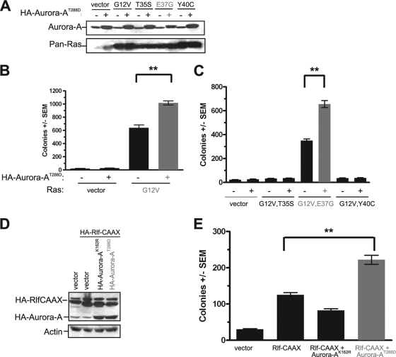
Aurora-AT288D promotes Ras-induced transformation through the RalGEF pathway. (A and D) Appropriate expression, as detected by immunoblot analysis, of kinase-active HA-Aurora-AT288D, kinase-inactive HA-Aurora-AK162R, constitutively activated HA-Rlf-CAAX, and ectopic and endogenous Ras (Pan-Ras). Actin serves as a loading control. (B, C, and E) Anchorage-independent growth in soft agar of polyclonal HEK-TtH cells stably expressing the indicated transgenes, expressed as average numbers of colonies formed ± standard errors of the means (SEM) for six plates (two independent experiments conducted in triplicate). The same vector (with [+] or without [−] HA-Aurora-AT288D) was used as a control in all of these experiments. Significant P values (<0.001) are indicated by **. Tukey's multiple-comparison test was used to determine significance between cell lines.
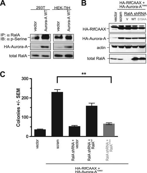
Aurora-A potentiates RalGEF-transformation through phosphorylation of RalA S194. (A) Appropriate expression, as detected by immunoblot analysis, of wild-type HA-Aurora-A, endogenous RalA, and phosphorylated RalA (detected by immunoprecipitation [IP] of endogenous RalA followed by immunoblot analysis [IB] with an anti-phospho-serine [α p-serine] antibody) in 293T and HEK-TtH cells expressing the indicated transgenes. Total RalA serves as a loading control. (B) Appropriate expression, as detected by immunoblot analysis, of HA-Aurora-AT288D, HA-Rlf-CAAX, knockdown of endogenous RalA, and complementation by ectopic RalA resistant to RalA shRNA in HEK-TtH cells stably infected with retroviruses carrying no transgene (vector [V]) or the indicated transgenes in the presence of shRNA specific to RalA or a scrambled version (scram) of this sequence. Actin serves as a loading control. (C) Anchorage-independent growth in soft agar of the aforementioned polyclonal HEK-TtH cells infected with retroviruses encoding the indicated shRNAs and carrying transgenes, expressed as average numbers of colonies formed ± SEM for six plates (two independent experiments conducted in triplicate). Significant P values (<0.001) are indicated by **. Tukey's multiple-comparison test was used to determine significance between cell lines.
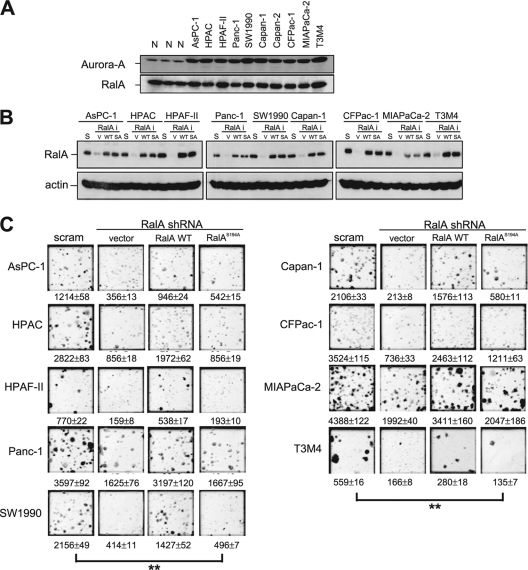
RalA S194 is broadly required for transformed growth of pancreatic cancer cell lines. (A) Detection of Aurora-A and RalA by immunoblot analysis of the indicated pancreatic cancer cell lines, compared with the results for three normal (N) pancreatic tissue specimens. (B) Immunoblot analysis of RalA in the indicated nine pancreatic cell lines, each retrovirally infected with a scramble sequence (S) or shRNA against RalA (RalAi) complemented by an empty vector (V), shRNA-resistant RalA in the wild-type (WT) or S194A mutant (SA) configuration. Actin serves as a loading control. (C) Photographs illustrating anchorage-independent growth in soft agar of the indicated polyclonal pancreatic cancer cells stably expressing either a RalA scramble sequence (scram) or RalA shRNA complemented with an empty vector (vector), the shRNA-resistant wild-type RalA protein (RalA WT), or the S194A mutant RalA protein (RalAS194A). Shown are average numbers of colonies formed ± SEM as calculated from triplicate plates. Data are from one representative experiment of two independent assays. Significant P values (<0.001) are indicated by **. Tukey's multiple-comparison test was used to determine significance between cell lines.
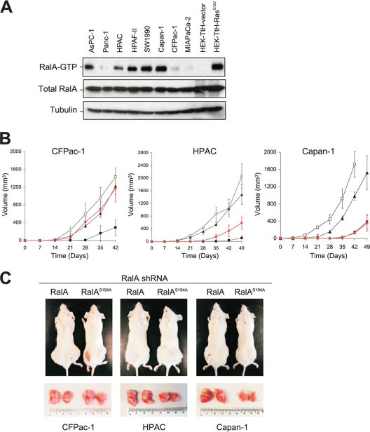
RalA S194 is required for tumorigenesis of pancreatic cancer cell lines. (A) RalA-GTP levels as detected in the indicated pancreatic cancer cell lines. HEK-TtH cells expressing empty vector or RasG12V serve as a negative or positive control, respectively. Total RalA and tubulin serve as loading controls. (B) Shown are tumor volumes (mm3) ± standard deviations versus times (days) observed for the indicated cell lines stably expressing a Ral scramble sequence (□), RalA-shRNA (▪), or RalA-shRNA complemented by expression of shRNA-resistant wild-type RalA (▴) or RalAS194A (•) injected into the flanks of immunocompromised mice. (C) Representative subcutaneous flank tumors observed in mice (top) and resected (bottom) from RalA shRNA-treated CFPac-1, HPAC, or Capan-1 cells transduced with shRNA-resistant wild-type RalA or the RalAS194A mutant at 42 days (CFPac-1) or 49 days (HPAC and Capan-1) after the cells were injected.
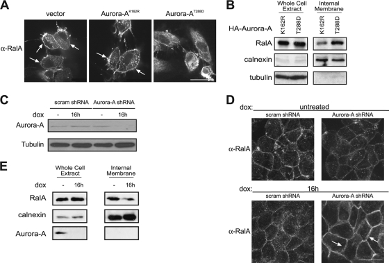
Aurora-A regulates RalA subcellular localization. (A) Immunofluorescence demonstrating distribution of endogenous RalA by use of an anti-RalA antibody in HEK-TtH cells stably expressing an empty vector, kinase-inactive HA (epitope-tagged)-Aurora-AK162R, or kinase-active HA-Aurora-AT288D. Arrows, plasma membrane localization. Scale bar, 20 μm. (B) Immunoblot analysis of endogenous RalA, calnexin, and tubulin in a whole-cell extract and an internal membrane fraction isolated from HEK-TtH cells expressing HA-Aurora-AK162R or HA-Aurora-A-T288D. (C) Reduction in endogenous Aurora-A protein, as detected by immunoblot analysis, in HPAC cells expressing doxycycline (dox)-inducible Aurora-A shRNA treated with dox for 16 h, compared to the level for untreated cells or dox-treated cells expressing a dox-inducible scramble control shRNA. Tubulin serves as a loading control. (D) Relocalization of endogenous RalA from the cytoplasm to the plasma membrane, as detected by immunofluorescence, in HPAC cells expressing dox-inducible Aurora-A shRNA treated with dox for 16 h, compared to the level for untreated cells or dox-treated cells expressing a dox-inducible scramble control shRNA. Arrows, plasma membrane localization. Scale bar, 20 μm. (E) Immunoblot analysis of endogenous RalA, calnexin, and tubulin in a whole-cell extract and an internal membrane fraction isolated from HPAC cells expressing dox-inducible Aurora-A shRNA treated with dox for 16 h, compared to the level for untreated cells.
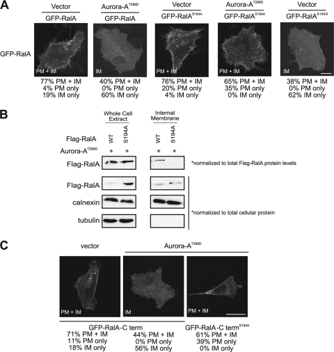
Aurora-AT288D-mediated internalization of RalA depends upon S194. (A) Distribution of GFP-RalA constructs in HEK-TtH cells stably expressing vector or HA-Aurora-AT288D in combination with GFP-RalA or GFP-RalAS194A, compared to the level for HEK-TtH cells expressing the phosphomimetic mutant RalA protein GFP-RalAS194D with vector alone. GFP-RalA localization to both the plasma membrane and the internal membrane (PM+IM), the plasma membrane only (PM), or the internal membrane only (IM) in 50 cells was quantitated in two independent experiments for each condition. Representative images with the primary location are displayed. Scale bar, 20 μm. (B) Immunoblot analysis of endogenous RalA, calnexin, and tubulin in a whole-cell extract and an internal membrane fraction isolated from HEK-TtH cells expressing HA-Aurora-AT288D with either wild-type or S194A Flag-RalA. Eightfold more RalAS194A than wild-type RalA was expressed in whole-cell extract. Therefore, wild-type Flag-RalA levels in the whole-cell extract and the internal membrane fraction were normalized 8:1 to S194A Flag-RalA levels. (C) Distribution of the last 20 amino acids of the RalA C terminus fused to GFP (GFP-RalA-C term) or, as indicated, GFP-RalA-C termS194A in HEK-TtH cells stably expressing either an empty vector, HA-Aurora-AK162R, or HA-Aurora-AT288D. GFP-RalA localization to both the plasma membrane and the internal membrane (PM+IM), the plasma membrane only (PM), or the internal membrane only (IM) in 50 cells was quantitated in two independent experiments for each condition. Representative images with the primary location are displayed. Scale bar, 20 μm.
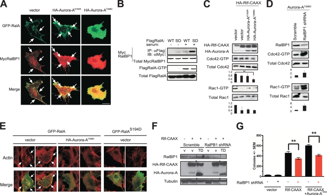
Aurora-A promotes cytoplasmic translocation and activation of RalBP1. (A) Distribution of GFP-RalA and Myc-tagged RalBP1 (MycRalBP1), visualized by immunofluorescence using an anti-Myc antibody in HEK-TtH cells stably expressing either a vector control, kinase-inactive HA-Aurora-AK162R, or kinase-active HA-Aurora-AT288D. Arrows, plasma membrane. Scale bar, 20 μm. (B) Aurora-A fosters the association of RalA with RalBP1. Immunoprecipitation (IP) of the Flag-tagged WT or the phosphomimetic S194D (SD) mutant version of RalA, followed by immunoblot analysis (IB) for detection of Flag-tagged, immunoprecipitated RalA protein or the presence or absence of coimmunoprecipitated Myc-tagged RalBP1 in the presence or absence of serum for activation of the RalA protein, as assessed by the level of Flag RalA-GTP. Total MycRalBP1 and Flag RalA serve as loading controls. (C) Aurora-A decreases Cdc42 and Rac1 activation. Shown are GTP-Cdc42 and GTP-Rac1 levels detected in HEK-TtH cells in which endogenous RalA was either not activated (vector) or activated by expressing HA-Rlf-CAAX in the presence, as assessed by immunoblot analysis, of either kinase-active HA-Aurora-AT288D or kinase-inactive HA-Aurora-AK162R. Total Cdc42 and Rac1 serve as loading controls. Cdc42-GTP and Rac1-GTP levels are normalized to levels for total Cdc42 and Rac1, expressed as fold changes ± standard deviations for three independent experiments (P = 0.029). (D) Knockdown of RalBP1 activates Cdc42 and Rac1. Shown are GTP-Cdc42 and GTP-Rac1 levels in HEK-TtH cells stably expressing shRNA against either the vector control or RalBP1. Total Cdc42 and Rac1 serve as loading controls. Cdc42-GTP and Rac1-GTP levels are normalized to the levels for total Cdc42 and Rac1, expressed as fold changes ± standard deviations for three independent experiments. (E) Distribution of GFP-RalA or GFP-RalAS194D and actin organization, visualized by immunofluorescence using Texas Red-phalloidin in HEK-TtH cells stably expressing either a vector control or kinase-active HA-Aurora-AT288D. Formation of filopodia and lamellipodia in 50 cells was quantitated in two independent experiments for each condition. Representative images are displayed. Arrows, filopodia or lamellipodia. Scale bar, 20 μm. (F) Appropriate expression, as detected by immunoblot analysis, of HA-Aurora-AT288D (TD), HA-Rlf-CAAX, or a knockdown of endogenous RalBP1 in HEK-TtH cells stably infected with retroviruses carrying no transgene (v) or the indicated transgenes in the presence of shRNA specific to RalBP1 or a scrambled version (scram) of this sequence. Tubulin serves as a loading control. (G) Anchorage-independent growth in soft agar of the aforementioned polyclonal HEK-TtH cells, infected with retroviruses encoding the indicated shRNAs and carrying the indicated transgenes, expressed as average numbers of colonies formed ± SEM for six plates (two independent experiments conducted in triplicate). Significant P values (<0.001) are indicated by **. Tukey's multiple-comparison test was used to determine significance between cell lines.
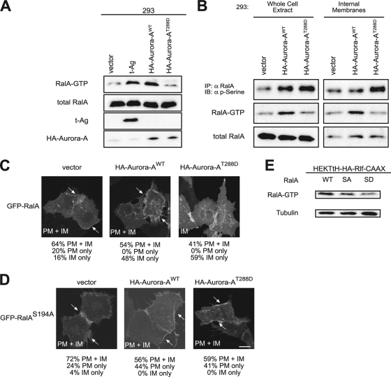
Differential effects of Aurora-A versus Aurora-AT288D on RalA functions. (A) Appropriate expression, as detected by immunoblot analysis, of t-Ag, wild-type HA-Aurora-A, kinase-active HA-Aurora-AT288D, endogenous RalA, and GTP-bound RalA levels (detected by GST pulldown of endogenous RalA) in 293 cells expressing the indicated transgenes. Total RalA serves as a loading control. (B) Immunoblot analysis of phosphorylated RalA (detected by immunoprecipitation of endogenous RalA followed by immunoblot analysis with an antiphosphoserine [α p-serine] antibody) and RalA GTP-levels (detected by GST pulldown of endogenous RalA) in a whole-cell extract and an internal membrane fraction isolated from 293 cells expressing the indicated transgenes. (C, D) Distribution of GFP-RalA or GFP-RalAS194A in HEK-TtH cells stably expressing vector, HA-Aurora-AWT, or HA-Aurora-AT288D. Arrows, plasma membrane localization. GFP-RalA localization to both the plasma membrane and the internal membrane (PM+IM), the plasma membrane only (PM), or the internal membrane only (IM) in 50 cells was quantitated in two independent experiments for each condition. Representative images with the primary location are displayed. Arrows, plasma membrane localization. Scale bar, 20 μm. (E) Immunoblot analysis of RalA-GTP levels in HEK-TtH cells stably expressing HA-Rlf-CAAX and wild-type RalA (WT), RalAS194A (SA), or RalAS194D (SD). Tubulin serves as a loading control.
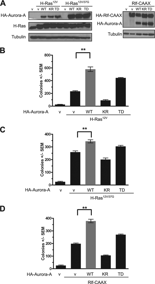
Wild-type versus kinase-active Aurora-A-mediated potentiation of Ras-induced transformation through the RalGEF pathway. (A) Appropriate expression, as detected by immunoblot analysis, of empty vector (v), wild-type HA-Aurora-A (WT), kinase-active HA-Aurora-AT288D (TD), kinase-inactive HA-Aurora-AK162R (KR), constitutively activated HA-Rlf-CAAX, or ectopic and endogenous Ras (Pan-Ras). Actin serves as a loading control. (B, C, D) Anchorage-independent growth in soft agar of polyclonal HEK-TtH cells stably expressing the indicated transgenes, expressed as average numbers of colonies formed ± SEM for six plates (two independent experiments conducted in triplicate). The same vector (v) (with [+] or without [−] HA-Aurora-A [WT], HA-Aurora-AT288D [TD], or HA-Aurora-AK162R [KR]) was used as a control in all of these experiments. Significant P values (<0.001) are indicated by **. Tukey's multiple-comparison test was used to determine significance between cell lines.
References
-
- Anand, S., S. Penrhyn-Lowe, and A. R. Venkitaraman. 2003. AURORA-A amplification overrides the mitotic spindle assembly checkpoint, inducing resistance to Taxol. Cancer Cell 3:51-62. - PubMed
-
- Awasthi, S., J. Cheng, S. S. Singhal, M. K. Saini, U. Pandya, S. Pikula, J. Bandorowicz-Pikula, S. V. Singh, P. Zimniak, and Y. C. Awasthi. 2000. Novel function of human RLIP76: ATP-dependent transport of glutathione conjugates and doxorubicin. Biochemistry 39:9327-9334. - PubMed
-
- Awasthi, S., S. S. Singhal, S. Yadav, J. Singhal, K. Drake, A. Nadkar, E. Zajac, D. Wickramarachchi, N. Rowe, A. Yacoub, P. Boor, S. Dwivedi, P. Dent, W. E. Jarman, B. John, and Y. C. Awasthi. 2005. RLIP76 is a major determinant of radiation sensitivity. Cancer Res. 65:6022-6028. - PubMed
-
- Bagrodia, S., S. J. Taylor, C. L. Creasy, J. Chernoff, and R. A. Cerione. 1995. Identification of a mouse p21Cdc42/Rac activated kinase. J. Biol. Chem. 270:22731-22737. - PubMed
-
- Bischoff, J. R., L. Anderson, Y. Zhu, K. Mossie, L. Ng, B. Souza, B. Schryver, P. Flanagan, F. Clairvoyant, C. Ginther, C. S. Chan, M. Novotny, D. J. Slamon, and G. D. Plowman. 1998. A homologue of Drosophila aurora kinase is oncogenic and amplified in human colorectal cancers. EMBO J. 17:3052-3065. - PMC - PubMed
Publication types
MeSH terms
Substances
Grants and funding
LinkOut - more resources
Full Text Sources
Molecular Biology Databases
Miscellaneous